


 申请试用
申请试用 产品介绍 评论(0)
宿主来源
Rabbit抗原名称
Galectin-3分子别名
Gal-3, 35 kDa lectin, CBP 35, Galactose-specific lectin 3, GALBP, IgE-binding protein, L-31, Laminin-binding protein, Lectin L-29, Mac-2 antigen免疫原
Recombinant Protein细胞定位
Cytoplasm, Nucleus, SecretedAccession
P17931克隆号
SDT-370-111抗体类型
Rabbit mAb应用
IHC-P ? ,ICC ? ,WB ,IP稀释度
应用 稀释度 WB 1:1000-1:5000 IHC-P 1:2500-1:5000 WB 1:500 IP 1:25 ICC 1:250 反应种属 ?
Hu纯化方式
Protein A浓度
1 mg/ml标记
Unconjugated性状
Liquid缓冲体系
PBS
储存条件
12 months from date of receipt, 4°C as supplied
Galectin-3 (Gal-3; formally named MAC-2) is a β-galactoside-binding lectin. Various cell types produce Gal-3 under either normal conditions and/or pathological conditions. Gal-3 can be present in cells' nuclei and cytoplasm, secreted from producing cells, and associated with cells' plasma membranes [PMID: 36274989].
免疫印迹
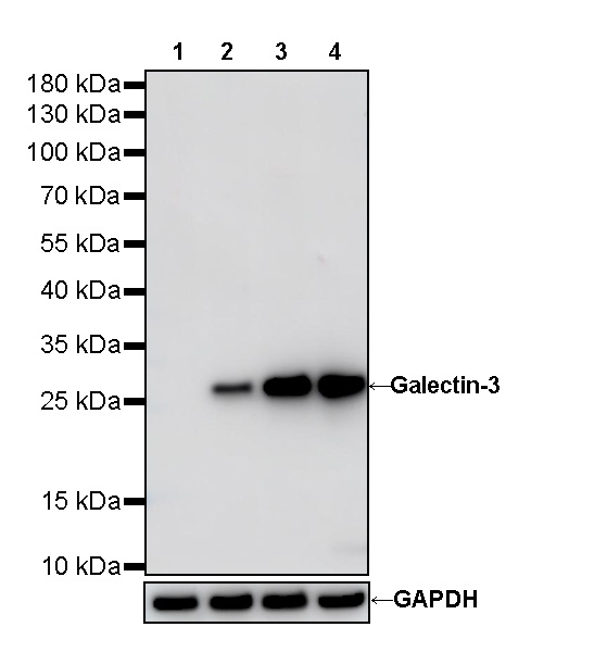
WB result of Galectin-3 Rabbit mAb
Primary antibody: Galectin-3 Rabbit mAb at 1/5000 dilution
Lane 1: LNCaP whole cell lysate 20 µg
Lane 2: HeLa whole cell lysate 20 µg
Lane 3: SW480 whole cell lysate 20 µg
Lane 4: MCF7 whole cell lysate 20 µg
Negative control: LNCaP whole cell lysate
Secondary antibody: Goat Anti-Rabbit IgG, (H+L), HRP conjugated at 1/10000 dilution
Predicted MW: 26 kDa
Observed MW: 28 kDa
免疫沉淀
Galectin-3 Rabbit mAb at 1/25 dilution (1 µg) immunoprecipitating Galectin-3 in 0.4 mg SW480 whole cell lysate.
Western blot was performed on the immunoprecipitate using Galectin-3 Rabbit mAb at 1/1000 dilution.
Secondary antibody (HRP) for IP was used at 1/400 dilution.
Lane 1: SW480 whole cell lysate 20 µg (Input)
Lane 2: Galectin-3 Rabbit mAb IP in SW480 whole cell lysate
Lane 3: Rabbit monoclonal IgG IP in SW480 whole cell lysate
Predicted MW: 26 kDa
Observed MW: 30 kDa
免疫组化
IHC shows positive staining in paraffin-embedded human colon cancer. Anti-Galectin-3 antibody was used at 1/2500 dilution, followed by a HRP Polymer for Mouse & Rabbit IgG (ready to use). Counterstained with hematoxylin. Heat mediated antigen retrieval with Tris/EDTA buffer pH9.0 was performed before commencing with IHC staining protocol.
IHC shows positive staining in paraffin-embedded human tonsil. Anti-Galectin-3 antibody was used at 1/2500 dilution, followed by a HRP Polymer for Mouse & Rabbit IgG (ready to use). Counterstained with hematoxylin. Heat mediated antigen retrieval with Tris/EDTA buffer pH9.0 was performed before commencing with IHC staining protocol.
IHC shows positive staining in paraffin-embedded human colon. Anti-Galectin-3 antibody was used at 1/2500 dilution, followed by a HRP Polymer for Mouse & Rabbit IgG (ready to use). Counterstained with hematoxylin. Heat mediated antigen retrieval with Tris/EDTA buffer pH9.0 was performed before commencing with IHC staining protocol.
Negative control: IHC shows negative staining in paraffin-embedded human renal clear cell carcinoma. Anti-Galectin-3 antibody was used at 1/2500 dilution, followed by a HRP Polymer for Mouse & Rabbit IgG (ready to use). Counterstained with hematoxylin. Heat mediated antigen retrieval with Tris/EDTA buffer pH9.0 was performed before commencing with IHC staining protocol.
Negative control: IHC shows negative staining in paraffin-embedded human papillary renal cell carcinoma. Anti-Galectin-3 antibody was used at 1/2500 dilution, followed by a HRP Polymer for Mouse & Rabbit IgG (ready to use). Counterstained with hematoxylin. Heat mediated antigen retrieval with Tris/EDTA buffer pH9.0 was performed before commencing with IHC staining protocol.
IHC shows positive staining in paraffin-embedded human chromophobe renal carcinoma. Anti-Galectin-3 antibody was used at 1/2500 dilution, followed by a HRP Polymer for Mouse & Rabbit IgG (ready to use). Counterstained with hematoxylin. Heat mediated antigen retrieval with Tris/EDTA buffer pH9.0 was performed before commencing with IHC staining protocol.
IHC shows positive staining in paraffin-embedded human oncocytic adenoma. Anti-Galectin-3 antibody was used at 1/2500 dilution, followed by a HRP Polymer for Mouse & Rabbit IgG (ready to use). Counterstained with hematoxylin. Heat mediated antigen retrieval with Tris/EDTA buffer pH9.0 was performed before commencing with IHC staining protocol.
IHC shows positive staining in paraffin-embedded human Hodgkin's lymphoma. Anti-Galectin-3 antibody was used at 1/5000 dilution, followed by a HRP Polymer for Mouse & Rabbit IgG (ready to use). Counterstained with hematoxylin. Heat mediated antigen retrieval with Tris/EDTA buffer pH9.0 was performed before commencing with IHC staining protocol.
Negative control: IHC shows negative staining in paraffin-embedded human follicular thyroid carcinoma. Anti-Galectin-3 antibody was used at 1/2500 dilution, followed by a HRP Polymer for Mouse & Rabbit IgG (ready to use). Counterstained with hematoxylin. Heat mediated antigen retrieval with Tris/EDTA buffer pH9.0 was performed before commencing with IHC staining protocol.
IHC shows positive staining in paraffin-embedded human papillary thyroid carcinoma. Anti-Galectin-3 antibody was used at 1/2500 dilution, followed by a HRP Polymer for Mouse & Rabbit IgG (ready to use). Counterstained with hematoxylin. Heat mediated antigen retrieval with Tris/EDTA buffer pH9.0 was performed before commencing with IHC staining protocol.
免疫细胞化学
ICC shows positive staining in MCF7 cells. Anti-Galectin-3 antibody was used at 1/250 dilution (Green) and incubated overnight at 4°C. Goat polyclonal Antibody to Rabbit IgG - H&L (Alexa Fluor® 488) was used as secondary antibody at 1/1000 dilution. The cells were fixed with 4% PFA and permeabilized with 0.1% PBS-Triton X-100. Nuclei were counterstained with DAPI (Blue). Counterstain with tubulin (red).
Negative control:ICC shows negative staining in LnCaP cells. Anti-Galectin-3 antibody was used at 1/250 dilution and incubated overnight at 4°C. Goat polyclonal Antibody to Rabbit IgG - H&L (Alexa Fluor® 488) was used as secondary antibody at 1/1000 dilution. The cells were fixed with 4% PFA and permeabilized with 0.1% PBS-Triton X-100. Nuclei were counterstained with DAPI (Blue). Counterstain with tubulin (red).

评论(0)