


 申请试用
申请试用 产品介绍 引用文献(2) 评论(0)
宿主来源
Rabbit抗原名称
CD3 epsilon分子别名
T-cell surface antigen T3/Leu-4 epsilon chain, CD3e, T3E, CD3E免疫原
Synthetic Peptide细胞定位
Cell membraneAccession
P07766克隆号
SDT-241-49抗体类型
Rabbit mAb反应种属 ?
Hu, Ms, Rt预测反应种属
(反应种属缩写表)Rb, Mq, Pg, Sh纯化方式
Protein A浓度
0.25 mg/ml性状
Liquid缓冲体系
PBS, 40% Glycerol, 0.05% BSA, 0.03% Proclin 300
储存条件
12 months from date of receipt / reconstitution, -20 °C as supplied
应用
稀释度
应用 稀释度 推荐种属 WB 1:1000-1:2000 Hu IHC-P 1:250-1:500 Hu, Ms, Rt IP 1:25 Hu ICC 1:250 Hu IF 1:1000 Hu
CD3 (cluster of differentiation 3) is a protein complex and T cell co-receptor that is involved in activating both the cytotoxic T cell (CD8+ naive T cells) and T helper cells (CD4+ naive T cells). It is composed of four distinct chains. In mammals, the complex contains a CD3γ chain, a CD3δ chain, and two CD3ε chains. These chains associate with the T-cell receptor (TCR) and the CD3-zeta (ζ-chain) to generate an activation signal in T lymphocytes. The TCR, CD3-zeta, and the other CD3 molecules together constitute the TCR complex. The CD3–T cell receptor (TCR) complex plays a central role in the T-cell-mediated immunoresponse as it is involved in the recognition of antigens and subsequent signal transduction and activation of immunocompetent T lymphocytes. Because CD3 is required for T cell activation, drugs (often monoclonal antibodies) that target it are being investigated as immunosuppressant therapies (e.g., otelixizumab, teplizumab) for type 1 diabetes and other autoimmune diseases.
免疫印迹
WB result of CD3 epsilon Rabbit mAb
Primary antibody: CD3 epsilon Rabbit mAb at 1/2000 dilution
Lane 1: Raji whole cell lysate 20 µg
Lane 2: Jurkat whole cell lysate 20 µg
Negative control: Raji whole cell lysate
Secondary antibody: Goat Anti-Rabbit IgG, (H+L), HRP conjugated at 1/10000 dilution
Predicted MW: 20 kDa
Observed MW: 20 kDa
免疫沉淀
CD3 epsilon Rabbit mAb at 1/25 dilution (1µg) immunoprecipitating CD3 epsilon in 0.4mg Jurkat whole cell lysate.
Western blot was performed on the immunoprecipitate using CD3 epsilon Rabbit mAb at 1/1000 dilution.
Secondary antibody (HRP) for IP was used at 1/400 dilution.
Lane 1 : Jurkat whole cell lysate 10µg(input)
Lane 2 : CD3 epsilon Rabbit mAb IP in Jurkat whole cell lysate
Lane 3 : Rabbit monoclonal IgG IP in Jurkat whole cell lysate
Predicted MW: 20 kDa
Observed MW: 20 kDa
免疫组化
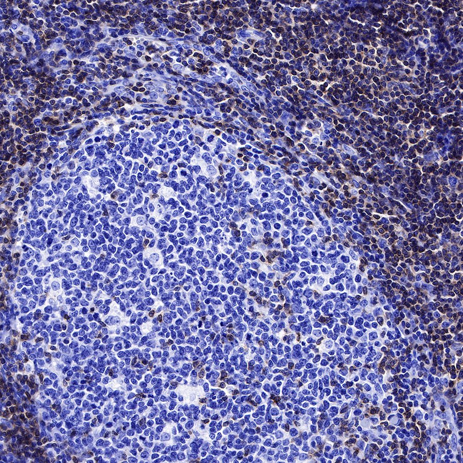
IHC shows positive staining in paraffin-embedded human spleen. Anti-CD3 epsilon antibody was used at 1/2000 dilution, followed by a HRP Polymer for Mouse & Rabbit IgG (ready to use). Counterstained with hematoxylin. Heat mediated antigen retrieval with Tris/EDTA buffer pH9.0 was performed before commencing with IHC staining protocol.
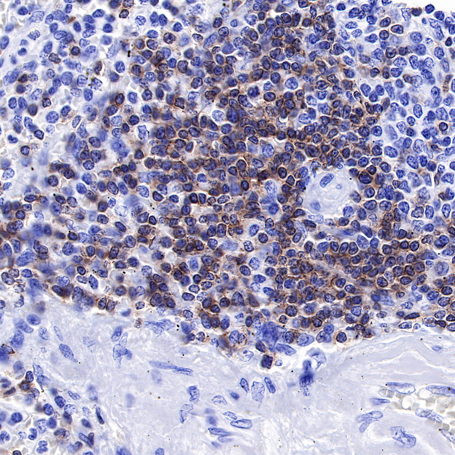
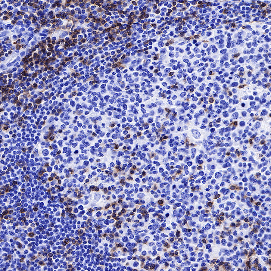
IHC shows positive staining in paraffin-embedded human tonsil. Anti-CD3 epsilon antibody was used at 1/500 dilution, followed by a HRP Polymer for Mouse & Rabbit IgG (ready to use). Counterstained with hematoxylin. Heat mediated antigen retrieval with Tris/EDTA buffer pH9.0 was performed before commencing with IHC staining protocol.
IHC shows positive staining in paraffin-embedded human tonsil. Anti-CD3 epsilon antibody was used at 1/500 dilution, followed by a HRP Polymer for Mouse & Rabbit IgG (ready to use). Counterstained with hematoxylin. Heat mediated antigen retrieval with Tris/EDTA buffer pH9.0 was performed before commencing with IHC staining protocol.
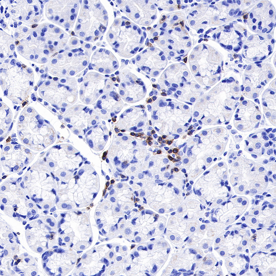
IHC shows positive staining in paraffin-embedded human stomach. Anti-CD3 epsilon antibody was used at 1/500 dilution, followed by a HRP Polymer for Mouse & Rabbit IgG (ready to use). Counterstained with hematoxylin. Heat mediated antigen retrieval with Tris/EDTA buffer pH9.0 was performed before commencing with IHC staining protocol.
IHC shows positive staining in paraffin-embedded mouse spleen. Anti-CD3 epsilon antibody was used at 1/500 dilution, followed by a HRP Polymer for Mouse & Rabbit IgG (ready to use). Counterstained with hematoxylin. Heat mediated antigen retrieval with Tris/EDTA buffer pH9.0 was performed before commencing with IHC staining protocol.
IHC shows positive staining in paraffin-embedded rat spleen. Anti-CD3 epsilon antibody was used at 1/500 dilution, followed by a HRP Polymer for Mouse & Rabbit IgG (ready to use). Counterstained with hematoxylin. Heat mediated antigen retrieval with Tris/EDTA buffer pH9.0 was performed before commencing with IHC staining protocol.
免疫细胞化学
ICC shows positive staining in Jurkat cells. Anti-CD3 epsilon antibody was used at 1/250 dilution (Green) and incubated overnight at 4°C. Goat polyclonal Antibody to Rabbit IgG - H&L (Alexa Fluor® 488) was used as secondary antibody at 1/1000 dilution. The cells were fixed with 4% PFA and permeabilized with 0.1% PBS-Triton X-100. Nuclei were counterstained with DAPI (Blue). Counterstain with tubulin (red).
免疫荧光
IF shows positive staining in paraffin-embedded human tonsil. Anti-CD3 epsilon antibody was used at 1/1000 dilution (Green) and incubated overnight at 4°C. Goat polyclonal Antibody to Rabbit IgG - H&L (Alexa Fluor® 488) was used as secondary antibody at 1/1000 dilution. Counterstained with DAPI (Blue). Heat mediated antigen retrieval with EDTA buffer pH9.0 was performed before commencing with IF staining protocol.
引用文献(2)
- Validation of CCL20-driven CAR-γδ T secreting PD-1 blockade with enhanced trafficking into solid tumor
Dengji Zhang, Yuan Tang, Wei Sun, ..., Xiaying Zhao, Xinyi Yang, Huanzhang Zhu
iScience. 45947 undefined undefined ; 10
影响因子: 4.1
货号:S0B2132产品名称:CD3 epsilon Recombinant Rabbit mAb (SDT-241-49)
- 人参皂苷F2对胆汁淤积肝损伤小鼠抗炎、抗氧化、抗纤维化和抗凋亡的作用机制
XU Huaming,YANG Liu,YAN Wuling,WANG Peiyu,ZHENG Sijia,LIU Yanxin,YANG Nian,ZHANG Xuelin,NIE Shanshan,FU Yinna,XING Weige,WANG Fuli
Drug Evaluation Research. 2025 Jan 09 .
影响因子: 2.434
货号:S0B2132产品名称:CD3 epsilon Recombinant Rabbit mAb (SDT-241-49)

评论(0)