


 申请试用
申请试用 产品介绍 FAQs 引用文献(4) 评论(0)
宿主来源
Rabbit抗原名称
F4/80分子别名
Adhesion G protein-coupled receptor E1, EGF-like module receptor 1, EMR1 hormone receptor, Gpf480, Adgre1免疫原
Synthetic Peptide细胞定位
Cell membraneAccession
Q61549克隆号
SDT-313-49抗体类型
Recombinant mAb应用
IHC-P ? ,ICC ? ,WB反应种属 ?
Ms, Rt纯化方式
Protein A浓度
0.5 mg/ml标记
Unconjugated性状
Liquid缓冲体系
PBS, 40% Glycerol, 0.05% BSA, 0.03% Proclin 300储存条件
12 months from date of receipt / reconstitution, -20 °C as supplied
| 应用 | 稀释度 |
|---|---|
| WB | 1:1000 |
| IHC-P | 1:500 |
| ICC | 1:100 |
The F4/80 monoclonal antibody has been used widely as a marker for mouse macrophages [PMID: 16087400]. EMR1-F4/80 is mainly present in the macrophage-containing stroma-vascular fraction. Furthermore, fibroblasts-like cells (adipoblasts), preadipocytes and adipocytes from the murine cell lines, 3T3-F442A and BFC-1, express CD14 and CD68 mRNA and protein as determined by fluorescence-activated cell sorter, but not F4/80 which, as expected, is strongly expressed in the macrophage cell line RAW264.7 [PMID: 16213494].
免疫印迹
WB result of F4/80 Rabbit mAb
Primary antibody: F4/80 Rabbit mAb at 1/1000 dilution
Lane 1: NIH/3T3 whole cell lysate 20 µg
Lane 2: RAW264.7 whole cell lysate 20 µg
Negative control: NIH/3T3 whole cell lysate
Secondary antibody: Goat Anti-Rabbit IgG, (H+L), HRP conjugated at 1/10000 dilution
Predicted MW: 97kDa
Observed MW: 300kDa
免疫组化
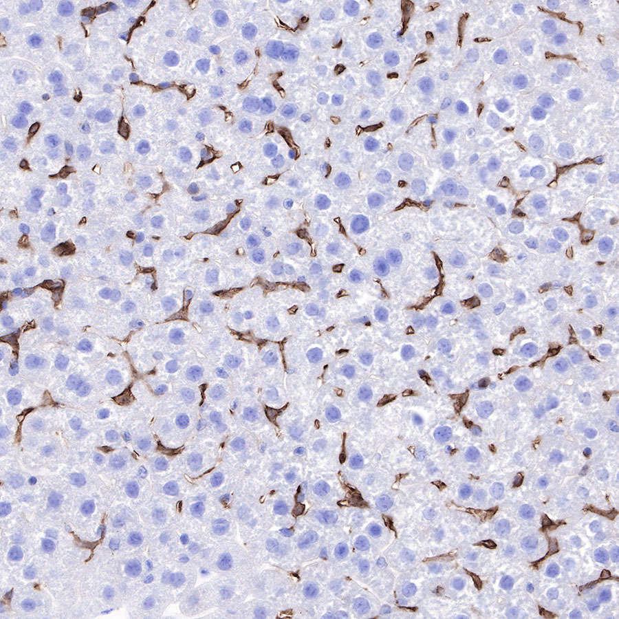
IHC shows positive staining in paraffin-embedded mouse liver. Anti-F4/80 antibody was used at 1/500 dilution, followed by a HRP Polymer for Mouse & Rabbit IgG (ready to use). Counterstained with hematoxylin. Heat mediated antigen retrieval with Tris/EDTA buffer pH9.0 was performed before commencing with IHC staining protocol.
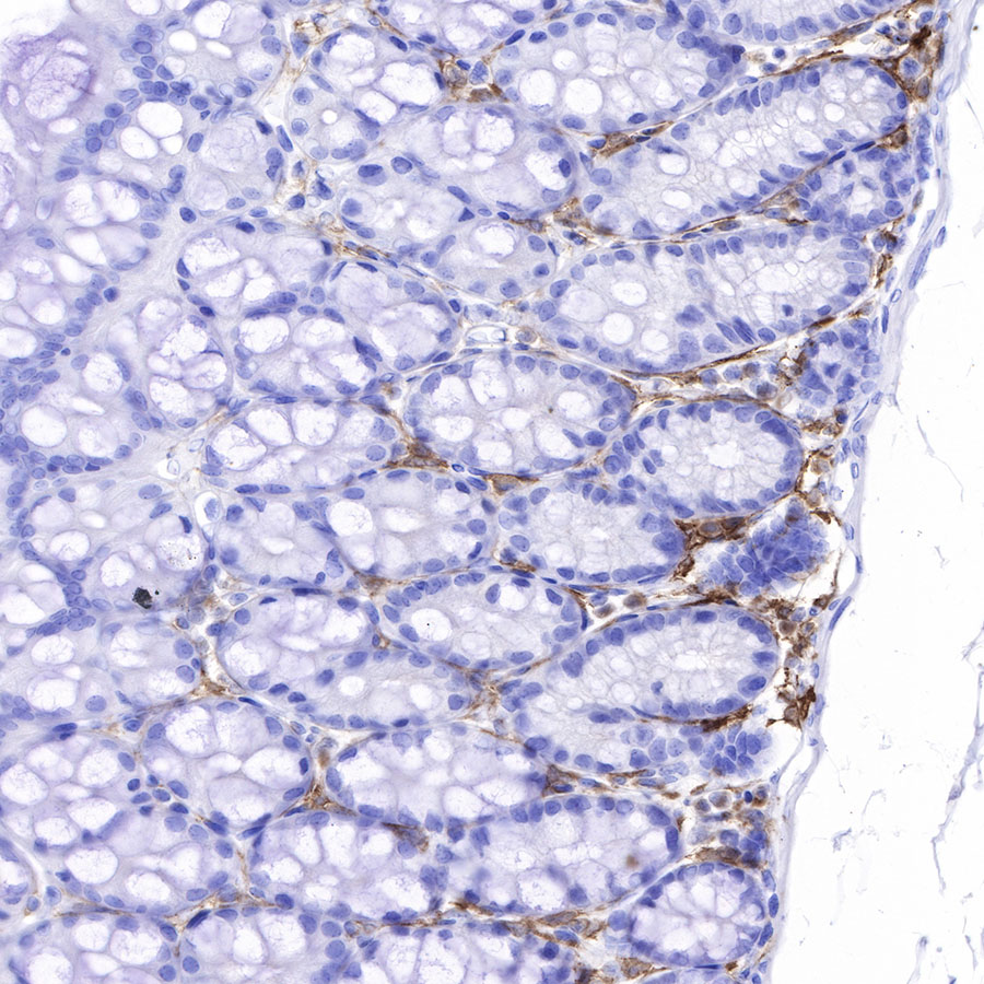
IHC shows positive staining in paraffin-embedded mouse colon. Anti-F4/80 antibody was used at 1/500 dilution, followed by a HRP Polymer for Mouse & Rabbit IgG (ready to use). Counterstained with hematoxylin. Heat mediated antigen retrieval with Tris/EDTA buffer pH9.0 was performed before commencing with IHC staining protocol.
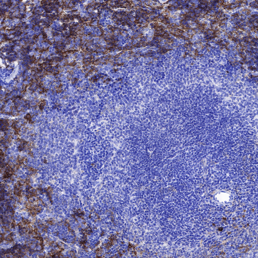
IHC shows positive staining in paraffin-embedded mouse spleen. Anti-F4/80 antibody was used at 1/500 dilution, followed by a HRP Polymer for Mouse & Rabbit IgG (ready to use). Counterstained with hematoxylin. Heat mediated antigen retrieval with Tris/EDTA buffer pH9.0 was performed before commencing with IHC staining protocol.
IHC shows positive staining in paraffin-embedded mouse stomach. Anti-F4/80 antibody was used at 1/500 dilution, followed by a HRP Polymer for Mouse & Rabbit IgG (ready to use). Counterstained with hematoxylin. Heat mediated antigen retrieval with Tris/EDTA buffer pH9.0 was performed before commencing with IHC staining protocol.
Negative control: IHC shows negative staining in paraffin-embedded mouse skeletal muscle. Anti-F4/80 antibody was used at 1/500 dilution, followed by a HRP Polymer for Mouse & Rabbit IgG (ready to use). Counterstained with hematoxylin. Heat mediated antigen retrieval with Tris/EDTA buffer pH9.0 was performed before commencing with IHC staining protocol.
IHC shows positive staining in paraffin-embedded rat liver. Anti-F4/80 antibody was used at 1/500 dilution, followed by a HRP Polymer for Mouse & Rabbit IgG (ready to use). Counterstained with hematoxylin. Heat mediated antigen retrieval with Tris/EDTA buffer pH9.0 was performed before commencing with IHC staining protocol.
免疫细胞化学
ICC shows positive staining in Raw264.7 cells. Anti-F4/80 antibody was used at 1/100 dilution (Green) and incubated overnight at 4°C. Goat polyclonal Antibody to Rabbit IgG - H&L (Alexa Fluor® 488) was used as secondary antibody at 1/1000 dilution. The cells were fixed with 100% ice-cold methanol and permeabilized with 0.1% PBS-Triton X-100. Nuclei were counterstained with DAPI (Blue).
Negative control:ICC shows negative staining in NIH/3T3 cells. Anti-F4/80 antibody was used at 1/100 dilution and incubated overnight at 4°C. Goat polyclonal Antibody to Rabbit IgG - H&L (Alexa Fluor® 488) was used as secondary antibody at 1/1000 dilution. The cells were fixed with 100% ice-cold methanol and permeabilized with 0.1% PBS-Triton X-100. Nuclei were counterstained with DAPI (Blue).
-
斯达特公司的抗体,可以回收利用几次?
引用文献(4)
- Peripheral nerve injury-induced remodeling of the tumor-associated macrophages promotes immune evasion in breast cancer
Y Jiang, W Zeng, Y Feng, X Deng, J Wang
Journal of Experimental & Clinical Cancer Research. 2025 Jan 01 .
影响因子: 11.4
货号:S0B0227产品名称:F4/80 Recombinant Rabbit mAb (SDT-313-49)
- Inhibition of Drp1- Fis1 interaction alleviates aberrant mitochondrial fragmentation and acute kidney injury
Z Song, Y Xia, L Shi, H Zha, J Huang, X Xiang
Cellular & Molecular Biology Letters. 2024 Mar 04 .
影响因子: 8.3
货号:S0B0227产品名称:F4/80 Recombinant Rabbit mAb (SDT-313-49)
- Inhibition of Drp1-mediated mitochondrial fission by P110 ameliorates renal injury in diabetic nephropathy
R Yue, Z Yan, H Zha, Y Xia, H Huang, H Li… - International …, 2025 - Elsevier
Int Immunopharmacol. 2025 Apr 16 .
影响因子: 4.7
货号:S0B0227产品名称:F4/80 Recombinant Rabbit mAb (SDT-313-49)
- Prostaglandin E2 alleviates inflammatory response and lung injury through EP4/cAMP/IKK/NF-κB pathway
Y Tang, W Pan, W Ding, X Pan, J Zhu, H Chen
Molecular Basis of Disease. 2025 Jan 01 ; 5
影响因子: 4.2
货号:S0B0227产品名称:F4/80 Recombinant Rabbit mAb (SDT-313-49)

评论(0)