


产品介绍 评论(0)
宿主来源
Rabbit抗原名称
VDAC1分子别名
Outer mitochondrial membrane protein porin 1; Plasmalemmal porin; Porin 31HL; Porin 31HM免疫原
Recombinant ProteinAccession
P21796克隆号
SDT-R013抗体类型
Rabbit mAb应用
IHC-P ? ,WB稀释度
应用 稀释度 推荐种属 IHC-P 1:2000 Hu, Ms, Rt WB 1:1000-1:10000 Hu, Ms, Rt, Zf 反应种属 ?
Hu, Ms, Rt, Zf纯化方式
Protein A浓度
0.5mg/ml分子量
32kDa
标记
Unconjugated性状
Liquid缓冲体系
PBS, 40% Glycerol, 0.05%BSA, 0.03% Proclin 300
储存条件
12 months from date of receipt / reconstitution, -20 °C as supplied.
Voltage-dependent anion-selective channel 1 (VDAC-1) is a beta barrel protein that in humans is encoded by the VDAC1 gene located on chromosome 5. It forms an ion channel in the outer mitochondrial membrane (OMM) and also the outer cell membrane. In the OMM, it allows ATP to diffuse out of the mitochondria into the cytoplasm. In the cell membrane, it is involved in volume regulation. Within all eukaryotic cells, mitochondria are responsible for synthesis of ATP among other metabolite needed for cell survival. VDAC1 therefore allows for communication between the mitochondrion and the cell mediating the balance between cell metabolism and cell death. Besides metabolic permeation, VDAC1 also acts as a scaffold for proteins such as hexokinase that can in turn regulate metabolism.
免疫印迹
WB result of VDAC1 Rabbit mAb
Primary antibody: VDAC1 Rabbit mAb at 1/1000 dilution
Lane 1: Hela whole cell lysate 20 µg
Lane 2: Jurkat whole cell lysate 20 µg
Secondary antibody: Goat Anti-Rabbit IgG, (H+L), HRP conjugated at 1/10000 dilution
Predicted MW: 32 kDa
Observed MW: 32 kDa
econdsWB result of VDAC1 Rabbit mAb
Primary antibody: VDAC1 Rabbit mAb at 1/1000 dilution
Lane 1: mouse kidney lysate 20 µg
Lane 2: NIH/3T3 whole cell lysate 20 µg
Secondary antibody: Goat Anti-Rabbit IgG, (H+L), HRP conjugated at 1/10000 dilution
Predicted MW: 32 kDa
Observed MW: 32 kDa
econdsWB result of VDAC1 Rabbit mAb
Primary antibody: VDAC1 Rabbit mAb at 1/1000 dilution
Lane 1: rat kidney lysate 20 µg
Secondary antibody: Goat Anti-Rabbit IgG, (H+L), HRP conjugated at 1/10000 dilution
Predicted MW: 32 kDa
Observed MW: 32 kDa
econdsWB result of VDAC1 Rabbit mAb
Primary antibody : VDAC1 Rabbit mAb at 1/1000 dilution
Lane 1 : Zebra fish lysate 20 µg
Secondary antibody: Goat Anti-Rabbit IgG, (H+L), HRP conjugated at 1/10000 dilution
Predicted MW: 32 kDa
Observed MW: 32 kDa
免疫组化
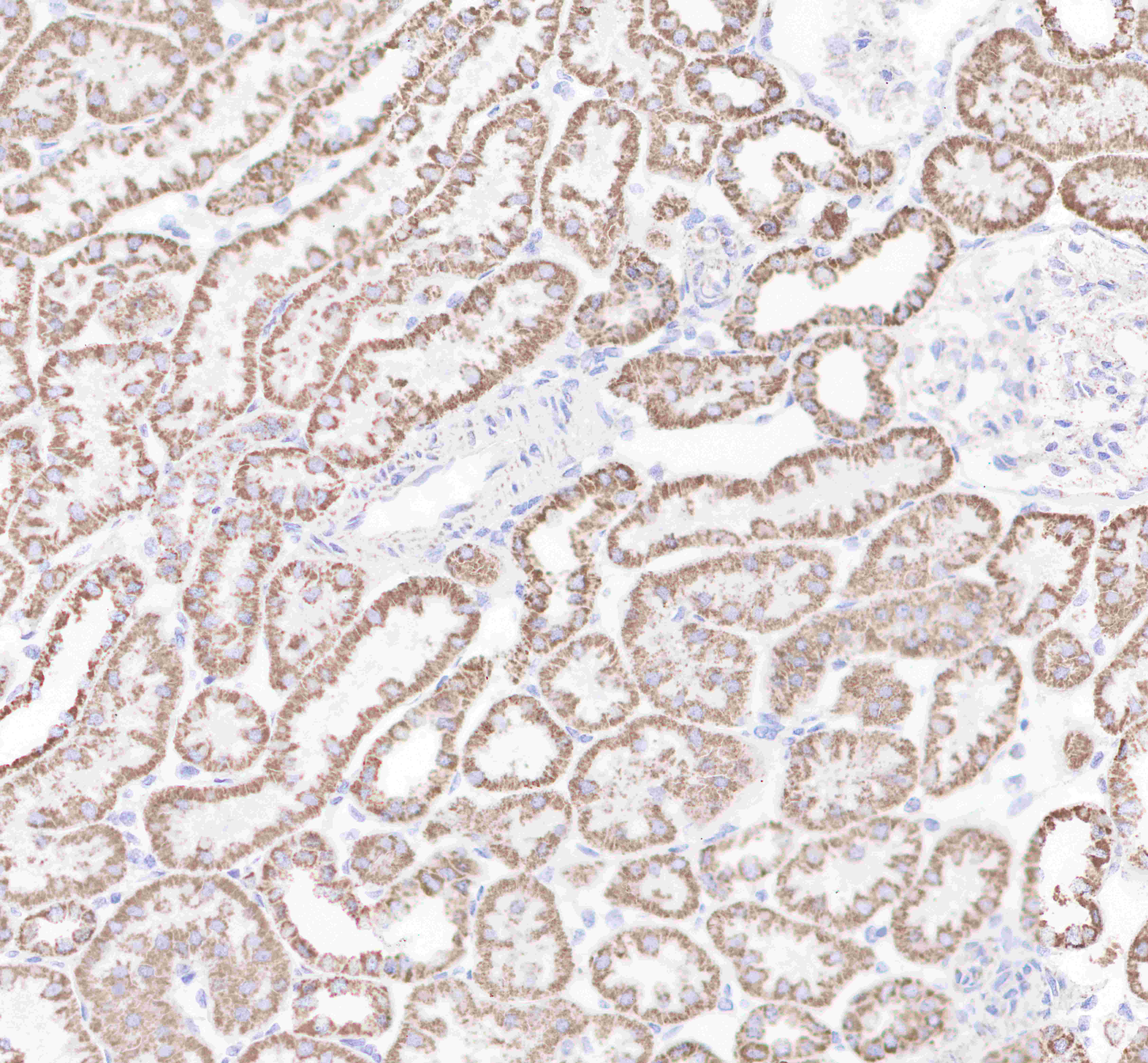
IHC shows positive staining in paraffin-embedded human kidney.
Anti-VDAC1 antibody was used at 1/2000 dilution, followed by a Goat Anti-Rabbit IgG H&L (HRP) ready to use.
Counterstained with hematoxylin.
Heat mediated antigen retrieval with Tris/EDTA buffer pH9.0 was performed before commencing with IHC staining protocol.
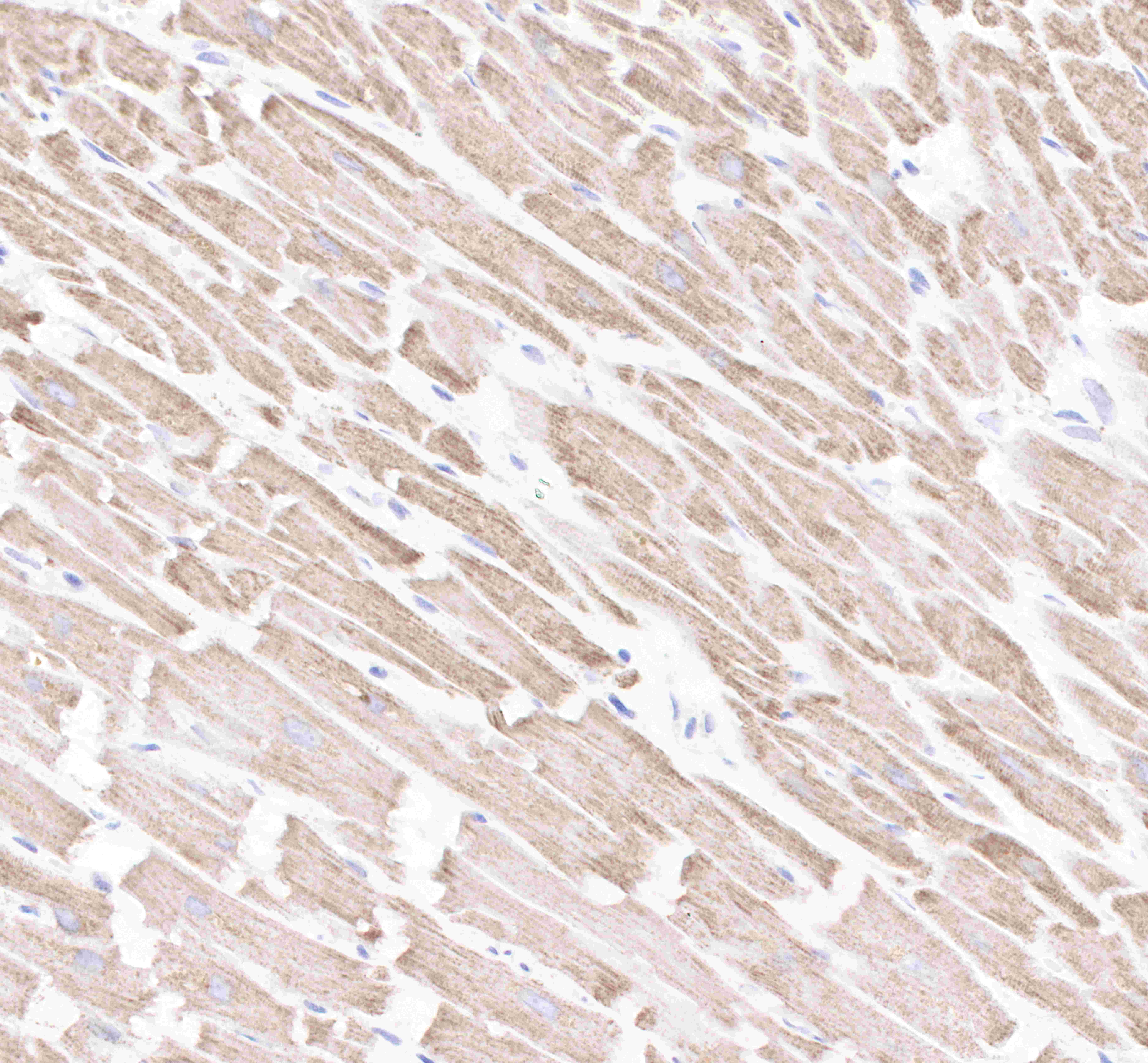
IHC shows positive staining in paraffin-embedded human myocardium.
Anti-VDAC1 antibody was used at 1/2000 dilution, followed by a Goat Anti-Rabbit IgG H&L (HRP) ready to use.
Counterstained with hematoxylin.
Heat mediated antigen retrieval with Tris/EDTA buffer pH9.0 was performed before commencing with IHC staining protocol.
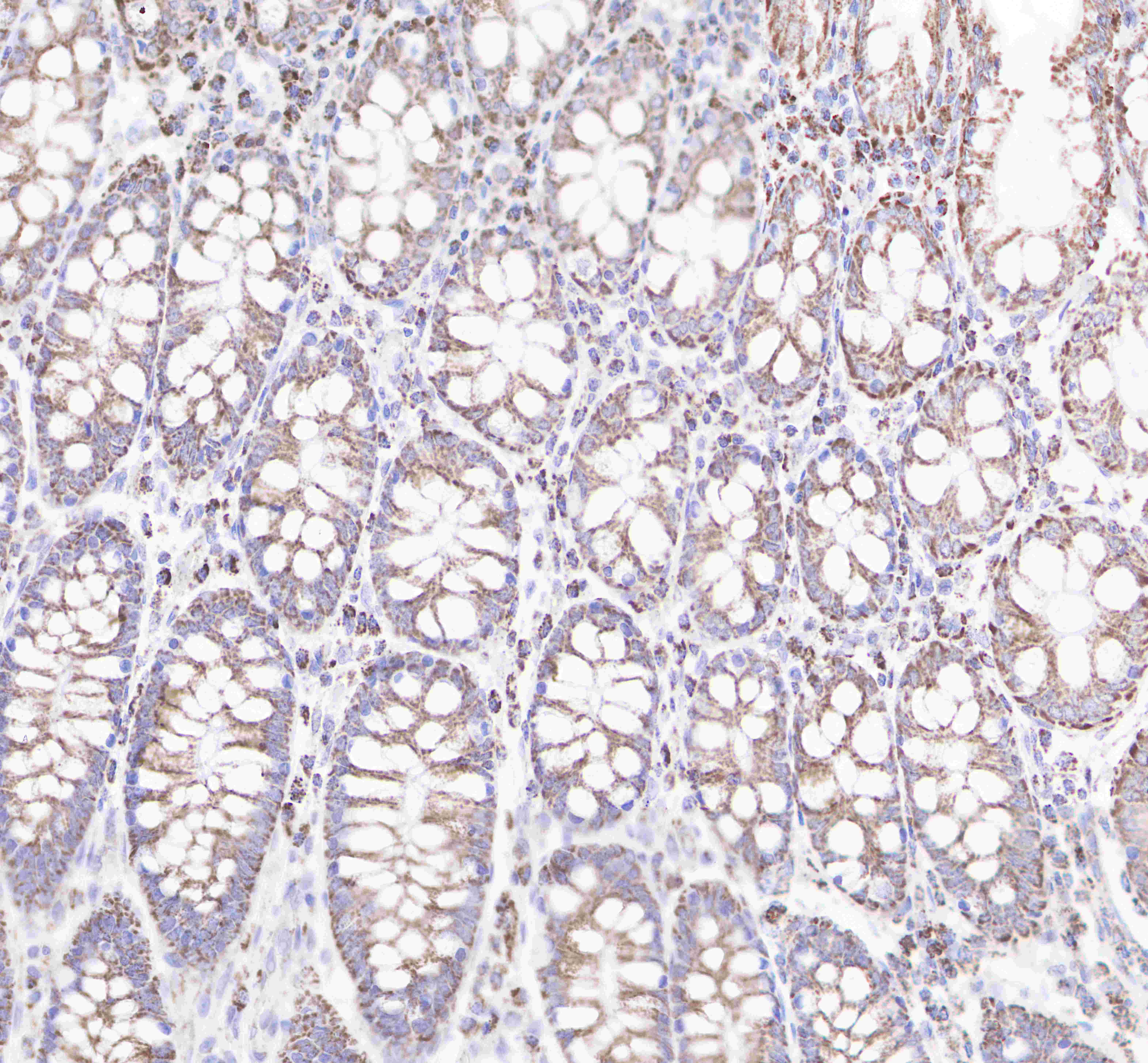
IHC shows positive staining in paraffin-embedded human colon.
Anti-VDAC1 antibody was used at 1/2000 dilution, followed by a Goat Anti-Rabbit IgG H&L (HRP) ready to use.
Counterstained with hematoxylin.
Heat mediated antigen retrieval with Tris/EDTA buffer pH9.0 was performed before commencing with IHC staining protocol.
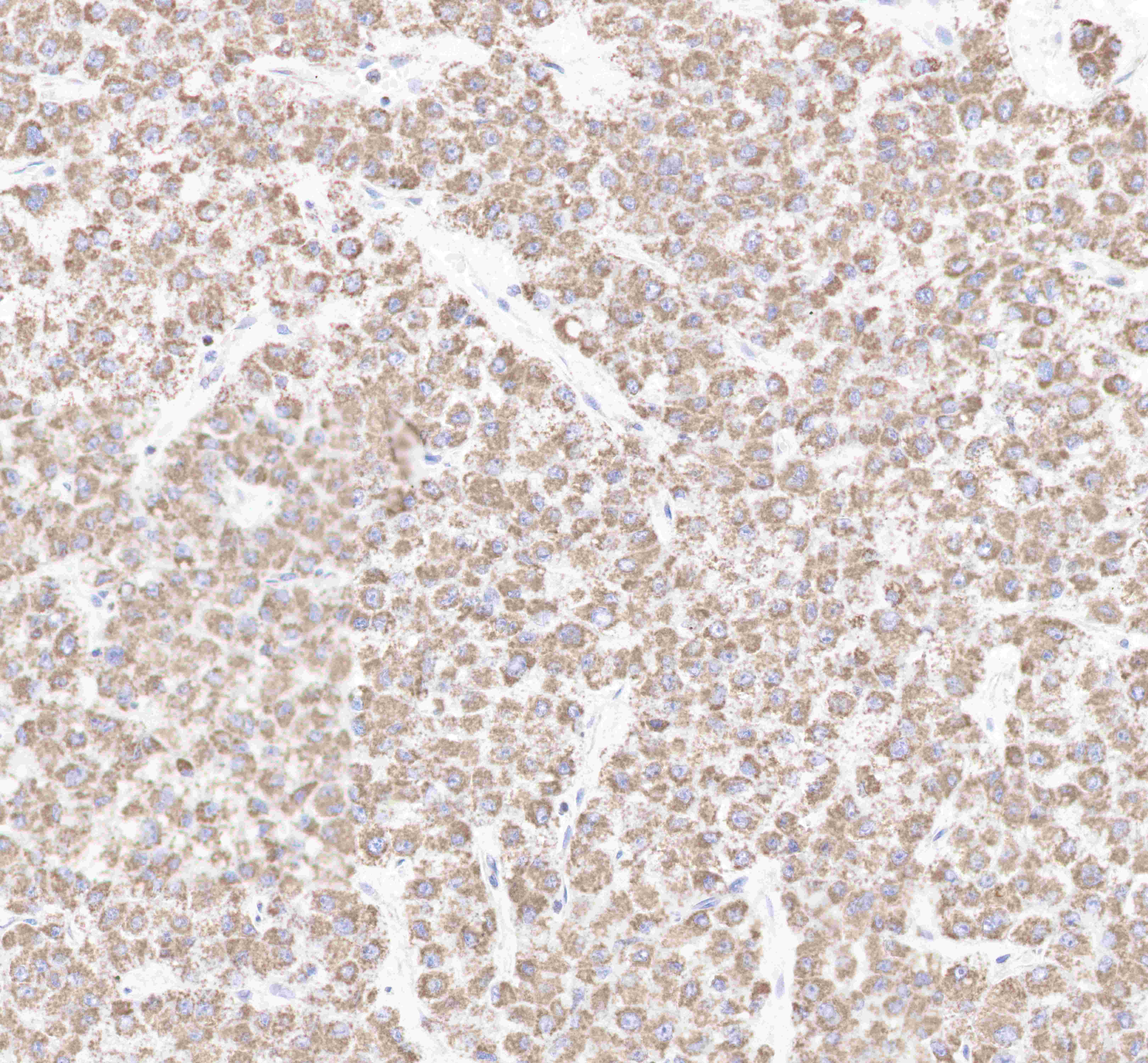
IHC shows positive staining in paraffin-embedded human hepatocellular carcinoma.
Anti-VDAC1 antibody was used at 1/2000 dilution, followed by a Goat Anti-Rabbit IgG H&L (HRP) ready to use.
Counterstained with hematoxylin.
Heat mediated antigen retrieval with Tris/EDTA buffer pH9.0 was performed before commencing with IHC staining protocol
IHC shows positive staining in paraffin-embedded human breast cancer.
Anti-VDAC1 antibody was used at 1/2000 dilution, followed by a Goat Anti-Rabbit IgG H&L (HRP) ready to use.
Counterstained with hematoxylin.
Heat mediated antigen retrieval with Tris/EDTA buffer pH9.0 was performed before commencing with IHC staining protocol.
IHC shows positive staining in paraffin-embedded mouse skeletal muscle.
Anti-VDAC1 antibody was used at 1/2000 dilution, followed by a Goat Anti-Rabbit IgG H&L (HRP) ready to use.
Counterstained with hematoxylin.
Heat mediated antigen retrieval with Tris/EDTA buffer pH9.0 was performed before commencing with IHC staining protocol.
IHC shows positive staining in paraffin-embedded rat kidney .
Anti-VDAC1 antibody was used at 1/2000 dilution, followed by a Goat Anti-Rabbit IgG H&L (HRP) ready to use.
Counterstained with hematoxylin.
Heat mediated antigen retrieval with Tris/EDTA buffer pH9.0 was performed before commencing with IHC staining protocol.

评论(0)