




 分享
分享产品介绍 FAQs 评论(0)
宿主来源
Rabbit抗原名称
CD10分子别名
Neprilysin, Atriopeptidase, Common acute lymphocytic leukemia antigen (CALLA), Enkephalinase, Neutral endopeptidase 24.11, NEP, Neutral endopeptidase, Skin fibroblast elastase (SFE), EPN免疫原
Synthetic Peptide细胞定位
Cell membraneAccession
P08473克隆号
SDT-006-77抗体类型
Recombinant mAb抗体同种型
IgG反应种属 ?
Hu, Ms, Rt纯化方式
Protein A浓度
1 mg/ml标记
Unconjugated性状
Liquid缓冲体系
PBS
储存条件
12 months from date of receipt / reconstitution, 4°C as supplied
应用
IHC-P ?
WB
IP
IF ?
稀释度
应用 稀释度 WB 1:1000 IHC 1:250 IF 1:100 IP 1:100
CD10, also known as membrane metallo-endopeptidase (MME), neutral endopeptidase (NEP), Neprilysin and common acute lymphoblastic leukemia antigen (CALLA) is an enzyme that in humans is encoded by the MME gene. Neprilysin is a zinc-dependent metalloprotease that cleaves peptides at the amino side of hydrophobic residues and inactivates several peptide hormones including glucagon, enkephalins, substance P, neurotensin, oxytocin, and bradykinin. It also degrades the amyloid beta peptide whose abnormal folding and aggregation in neural tissue has been implicated as a cause of Alzheimer's disease. Neprilysin is expressed in a wide variety of tissues and is particularly abundant in kidney. It is also a common acute lymphocytic leukemia antigen that is an important cell surface marker in the diagnosis of human acute lymphocytic leukemia (ALL). This protein is present on leukemic cells of pre-B phenotype, which represent 85% of cases of ALL. Hematopoietic progenitors expressing CD10 are considered "common lymphoid progenitors", which means they can differentiate into T, B or natural killer cells. CD10 is of use in hematological diagnosis since it is expressed by early B, pro-B and pre-B lymphocytes, and by lymph node germinal centers. Hematologic diseases in which it is positive include ALL, angioimmunoblastic T cell lymphoma, Burkitt lymphoma, chronic myelogenous leukemia in blast crisis (90%), diffuse large B-cell lymphoma (variable), follicular center cells (70%), hairy cell leukemia (10%), and myeloma (some). It tends to be negative in acute myeloid leukemia, chronic lymphocytic leukemia, mantle cell lymphoma, and marginal zone lymphoma.
免疫印迹
WB result of CD10 Rabbit mAb
Primary antibody: CD10 Rabbit mAb at 1/1000 dilution
Lane 1: Jurkat whole cell lysate 20 µg
Lane 2: Raji whole cell lysate 20 µg
Lane 3: Ramos whole cell lysate 20 µg
Negative control: Jurkat whole cell lysate
Secondary antibody: Goat Anti-Rabbit IgG, (H+L), HRP conjugated at 1/10000 dilution
Predicted MW: 86 kDa
Observed MW: 110 kDaWB result of CD10 Rabbit mAb
Primary antibody: CD10 Rabbit mAb at 1/1000 dilution
Lane 1: mouse kidney lysate 20 µg
Secondary antibody: Goat Anti-Rabbit IgG, (H+L), HRP conjugated at 1/10000 dilution
Predicted MW: 86 kDa
Observed MW: 110 kDaWB result of CD10 Rabbit mAb
Primary antibody: CD10 Rabbit mAb at 1/1000 dilution
Lane 1: rat kidney lysate 20 µg
Secondary antibody: Goat Anti-Rabbit IgG, (H+L), HRP conjugated at 1/10000 dilution
Predicted MW: 86 kDa
Observed MW: 110 kDa
免疫沉淀
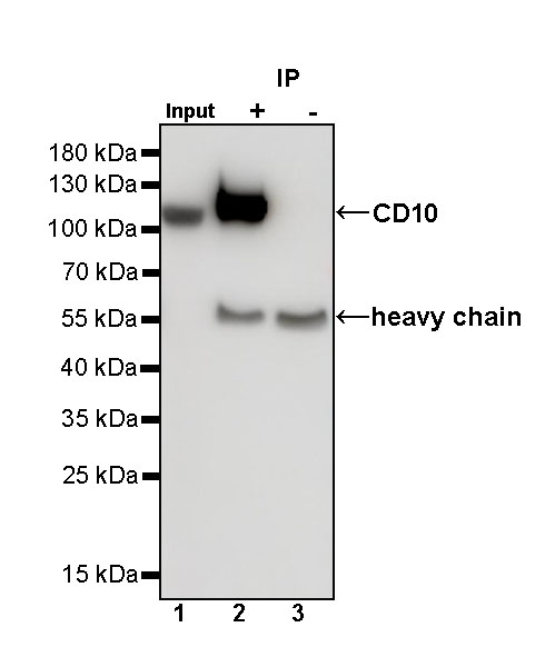
CD10 Rabbit mAb at 1/100 dilution (0.8 µg) immunoprecipitating CD10 in 0.4 mg Ramos whole cell lysate.
Western blot was performed on the immunoprecipitate using CD10 Rabbit mAb at 1/1000 dilution.
Secondary antibody (HRP) for IP was used at 1/400 dilution.
Lane 1: Ramos whole cell lysate 20 µg (Input)
Lane 2: CD10 Rabbit mAb IP in Ramos whole cell lysate
Lane 3: Rabbit monoclonal IgG IP in Ramos whole cell lysate
Predicted MW: 86 kDa
Observed MW: 110 kDa
免疫组化
IHC shows positive staining in paraffin-embedded human kidney. Anti-CD10 antibody was used at 1/250 dilution, followed by a HRP Polymer for Mouse & Rabbit IgG (ready to use). Counterstained with hematoxylin. Heat mediated antigen retrieval with Tris/EDTA buffer pH9.0 was performed before commencing with IHC staining protocol.
IHC shows positive staining in paraffin-embedded human liver. Anti-CD10 antibody was used at 1/250 dilution, followed by a HRP Polymer for Mouse & Rabbit IgG (ready to use). Counterstained with hematoxylin. Heat mediated antigen retrieval with Tris/EDTA buffer pH9.0 was performed before commencing with IHC staining protocol.
IHC shows positive staining in paraffin-embedded mouse kidney. Anti-CD10 antibody was used at 1/250 dilution, followed by a HRP Polymer for Mouse & Rabbit IgG (ready to use). Counterstained with hematoxylin. Heat mediated antigen retrieval with Tris/EDTA buffer pH9.0 was performed before commencing with IHC staining protocol.
免疫荧光
IF shows positive staining in paraffin-embedded human tonsil. Anti-CD10 antibody was used at 1/100 dilution (Green) and incubated overnight at 4°C. Goat polyclonal Antibody to Rabbit IgG - H&L (Alexa Fluor® 488) was used as secondary antibody at 1/1000 dilution. Counterstained with DAPI (Blue). Heat mediated antigen retrieval with EDTA buffer pH9.0 was performed before commencing with IF staining protocol.
IF shows positive staining in paraffin-embedded human kidney. Anti-CD10 antibody was used at 1/100 dilution (Green) and incubated overnight at 4°C. Goat polyclonal Antibody to Rabbit IgG - H&L (Alexa Fluor® 488) was used as secondary antibody at 1/1000 dilution. Counterstained with DAPI (Blue). Heat mediated antigen retrieval with EDTA buffer pH9.0 was performed before commencing with IF staining protocol.
FAQs
这个克隆在扁桃体样本上表现的强度较弱,不推荐在扁桃体样本上使用?

评论(0)