


p40(BC28)Recombinant Rabbit mAb,PBS Only (SDT-166-36)
Tumor protein 63,p63,Chronic ulcerative stomatitis protein (CUSP),Keratinocyte transcription factor KET,Transformation-related protein 63 (TP63),Tumor protein p73-like (p73L),p51,TP63,KET,P73H,P73L,TP73L


 分享
分享产品介绍 评论(0)
宿主来源
Rabbit抗原名称
p40分子别名
Tumor protein 63; p63; Chronic ulcerative stomatitis protein (CUSP); Keratinocyte transcription factor KET; Transformation-related protein 63 (TP63); Tumor protein p73-like (p73L); p51; TP63; KET; P73H; P73L; TP73L免疫原
Synthetic Peptide细胞定位
NucleusAccession
Q9H3D4-4克隆号
SDT-166-36抗体类型
Recombinant mAb反应种属 ?
Hu纯化方式
Protein A浓度
1 mg/ml标记
Unconjugated性状
Liquid缓冲体系
PBS
储存条件
12 months from date of receipt, 4°C as supplied
应用
IHC-P ?
WB
IP
稀释度
应用 稀释度 WB 1:1000 IP 1:50 IHC-P 1:500
p63 (TAp63) is closely related to p40 (ΔNp63) as both proteins represent isoforms of the p63 gene with distinct molecular functions. While “full length” p63 (TAp63) activates p53 target genes such as p21 or BAX, the shorter transcript p40 (ΔNp63) inhibits activation of p53 and “full length” p63. Among carcinomas, p40 has approximately the same sensitivity as p63 but a higher specificity, as the TAp63 isoform is expressed more widespread in eg., adenocarcinomas. Moreover, p63 occur in lymphomas that are p40 negative.
免疫印迹
WB result of p40 Rabbit mAb Primary antibody: p40 Rabbit mAb at 1/1000 dilution Lane 1: MCF7 whole cell lysate 20 µg Lane 2: A431 whole cell lysate 20 µg Lane 3: HaCaT whole cell lysate 20 µg Negative control: MCF7 whole cell lysate Secondary antibody: Goat Anti-Rabbit IgG, (H+L), HRP conjugated at 1/10000 dilution Predicted MW: 51 kDa Observed MW: 51, 75 kDa
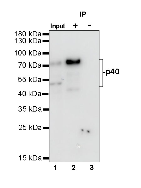
免疫沉淀
p40 Rabbit mAb at 1/50 dilution (1 µg) immunoprecipitating p40 in 0.4 mg HaCaT whole cell lysate.
Western blot was performed on the immunoprecipitate using p40 Rabbit mAb at 1/1000 dilution.
Secondary antibody (HRP) for IP was used at 1/400 dilution.
Lane 1: HaCaT whole cell lysate 10 µg (Input)
Lane 2: p40 Rabbit mAb IP in HaCaT whole cell lysate
Lane 3: Rabbit monoclonal IgG IP in HaCaT whole cell lysate
Predicted MW: 51 kDa
Observed MW: 51, 75 kDa
(This blot was developed with high sensitivity substrate)
免疫组化
Negative control: IHC shows negative staining in paraffin-embedded human diffuse large B-cell lymphoma. Anti-p40 antibody was used at 1/500 dilution, followed by a HRP Polymer for Mouse & Rabbit IgG (ready to use). Counterstained with hematoxylin. Heat mediated antigen retrieval with Tris/EDTA buffer pH9.0 was performed before commencing with IHC staining protocol.
Negative control: IHC shows negative staining in paraffin-embedded human lung adenocarcinoma. Anti-p40 antibody was used at 1/500 dilution, followed by a HRP Polymer for Mouse & Rabbit IgG (ready to use). Counterstained with hematoxylin. Heat mediated antigen retrieval with Tris/EDTA buffer pH9.0 was performed before commencing with IHC staining protocol.
IHC shows positive staining in paraffin-embedded human lung squamous cell carcinoma. Anti-p40 antibody was used at 1/500 dilution, followed by a HRP Polymer for Mouse & Rabbit IgG (ready to use). Counterstained with hematoxylin. Heat mediated antigen retrieval with Tris/EDTA buffer pH9.0 was performed before commencing with IHC staining protocol.
IHC shows positive staining in paraffin-embedded human breast cancer. Anti-p40 antibody was used at 1/500 dilution, followed by a HRP Polymer for Mouse & Rabbit IgG (ready to use). Counterstained with hematoxylin. Heat mediated antigen retrieval with Tris/EDTA buffer pH9.0 was performed before commencing with IHC staining protocol.
IHC shows positive staining in paraffin-embedded human tonsil. Anti-p40 antibody was used at 1/500 dilution, followed by a HRP Polymer for Mouse & Rabbit IgG (ready to use). Counterstained with hematoxylin. Heat mediated antigen retrieval with Tris/EDTA buffer pH9.0 was performed before commencing with IHC staining protocol.

评论(0)