


 大包装询价
大包装询价 产品介绍 评论(0)
宿主来源
Rabbit抗原名称
CD79a/MB-1分子别名
Ig-alpha, Surface IgM-associated protein, IGA免疫原
Synthetic Peptide细胞定位
Cell membraneAccession
P11912克隆号
SDT-030-18抗体类型
Recombinant mAb应用
IHC-P, WB, IF反应种属 ?
Hu纯化方式
Protein A浓度
1 mg/ml标记
Unconjugated性状
Liquid缓冲体系
PBS储存条件
12 months from date of receipt, 4°C as supplied
| 应用 | 稀释度 |
|---|---|
| WB | 1:5000 |
| IHC-P | 1:1000 |
| IF | 1:500 |
The CD79 molecule, comprising two polypeptide chains, mb-1 (CD79a) and B29 (CD79b), is physically associated in the B-cell membrane with immunoglobulin [PMID: 7632952]. CD79a has been reported to exhibit a high degree of linage-specificity for B-cell differentiation, with a specificity of 88% and a sensitivity of 100% [PMID: 34672246].
免疫印迹
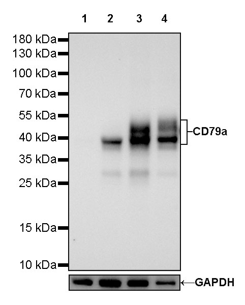
WB result of CD79a Rabbit mAb
Primary antibody: CD79a Rabbit mAb at 1/5000 dilution
Lane 1: Jurkat whole cell lysate 20 µg
Lane 2: Raji whole cell lysate 20 µg
Lane 3: Ramos whole cell lysate 20 µg
Lane 4: Daudi whole cell lysate 20 µg
Negative control: Jurkat whole cell lysate
Secondary antibody: Goat Anti-Rabbit IgG, (H+L), HRP conjugated at 1/10000 dilution
Predicted MW: 25kDa
Observed MW: 40~55kDa
免疫组化

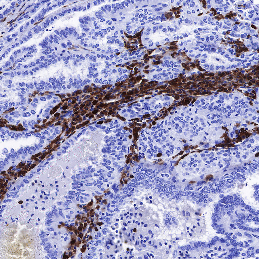
IHC shows positive staining in paraffin-embedded human colon. Anti-CD79a/MB-1 antibody was used at 1/1000 dilution, followed by a HRP Polymer for Mouse & Rabbit IgG (ready to use). Counterstained with hematoxylin. Heat mediated antigen retrieval with Tris/EDTA buffer pH9.0 was performed before commencing with IHC staining protocol.

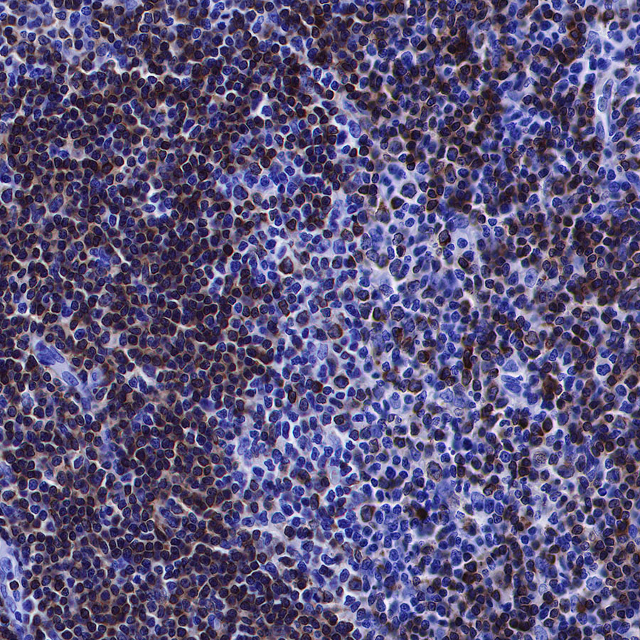
IHC shows positive staining in paraffin-embedded human spleen. Anti-CD79a/MB-1 antibody was used at 1/1000 dilution, followed by a HRP Polymer for Mouse & Rabbit IgG (ready to use). Counterstained with hematoxylin. Heat mediated antigen retrieval with Tris/EDTA buffer pH9.0 was performed before commencing with IHC staining protocol.
IHC shows positive staining in paraffin-embedded human tonsil. Anti-CD79a/MB-1 antibody was used at 1/1000 dilution, followed by a HRP Polymer for Mouse & Rabbit IgG (ready to use). Counterstained with hematoxylin. Heat mediated antigen retrieval with Tris/EDTA buffer pH9.0 was performed before commencing with IHC staining protocol.
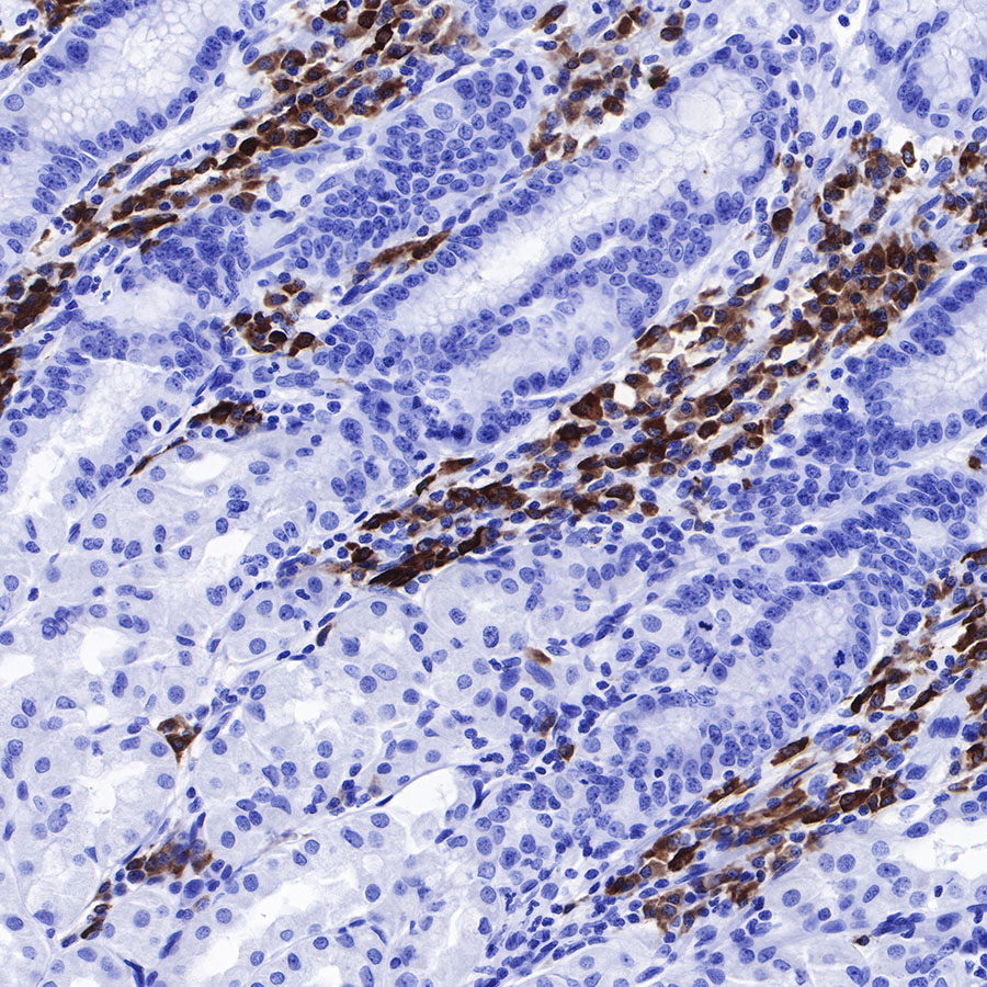
IHC shows positive staining in paraffin-embedded human stomach. Anti-CD79a/MB-1 antibody was used at 1/1000 dilution, followed by a HRP Polymer for Mouse & Rabbit IgG (ready to use). Counterstained with hematoxylin. Heat mediated antigen retrieval with Tris/EDTA buffer pH9.0 was performed before commencing with IHC staining protocol.
IHC shows positive staining in paraffin-embedded human thyroid cancer. Anti-CD79a/MB-1 antibody was used at 1/1000 dilution, followed by a HRP Polymer for Mouse & Rabbit IgG (ready to use). Counterstained with hematoxylin. Heat mediated antigen retrieval with Tris/EDTA buffer pH9.0 was performed before commencing with IHC staining protocol.
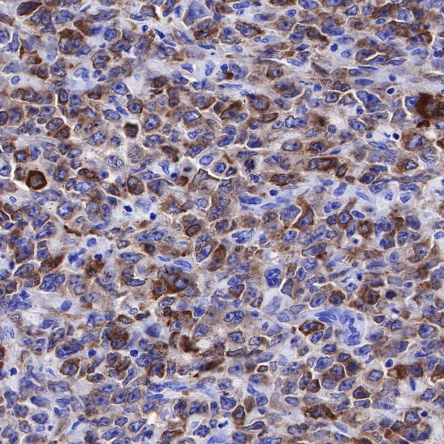
IHC shows positive staining in paraffin-embedded human diffuse large B-cell lymphoma. Anti-CD79a/MB-1 antibody was used at 1/1000 dilution, followed by a HRP Polymer for Mouse & Rabbit IgG (ready to use). Counterstained with hematoxylin. Heat mediated antigen retrieval with Tris/EDTA buffer pH9.0 was performed before commencing with IHC staining protocol.

Negative control: IHC shows negative staining in paraffin-embedded human skeletal muscle. Anti-CD79a/MB-1 antibody was used at 1/1000 dilution, followed by a HRP Polymer for Mouse & Rabbit IgG (ready to use). Counterstained with hematoxylin. Heat mediated antigen retrieval with Tris/EDTA buffer pH9.0 was performed before commencing with IHC staining protocol.
免疫荧光
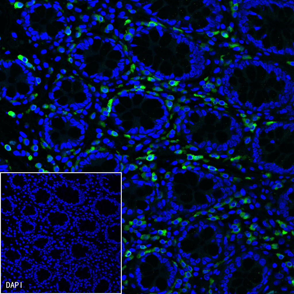
IF shows positive staining in paraffin-embedded human colon. Anti-CD79a/MB-1 antibody was used at 1/500 dilution (Green) and incubated overnight at 4°C. Goat polyclonal Antibody to Rabbit IgG - H&L (Alexa Fluor® 488) was used as secondary antibody at 1/1000 dilution. Counterstained with DAPI (Blue). Heat mediated antigen retrieval with EDTA buffer pH9.0 was performed before commencing with IF staining protocol.
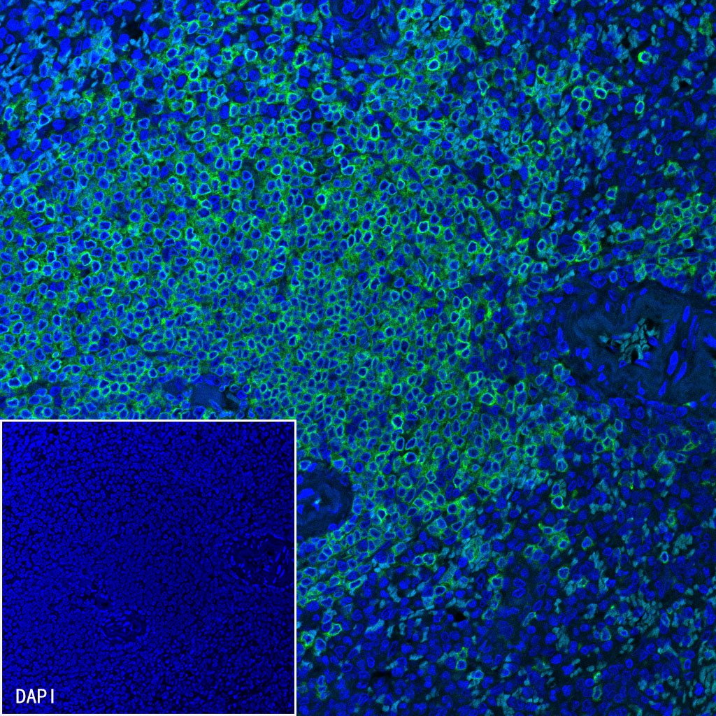
IF shows positive staining in paraffin-embedded human spleen. Anti-CD79a/MB-1 antibody was used at 1/500 dilution (Green) and incubated overnight at 4°C. Goat polyclonal Antibody to Rabbit IgG - H&L (Alexa Fluor® 488) was used as secondary antibody at 1/1000 dilution. Counterstained with DAPI (Blue). Heat mediated antigen retrieval with EDTA buffer pH9.0 was performed before commencing with IF staining protocol.
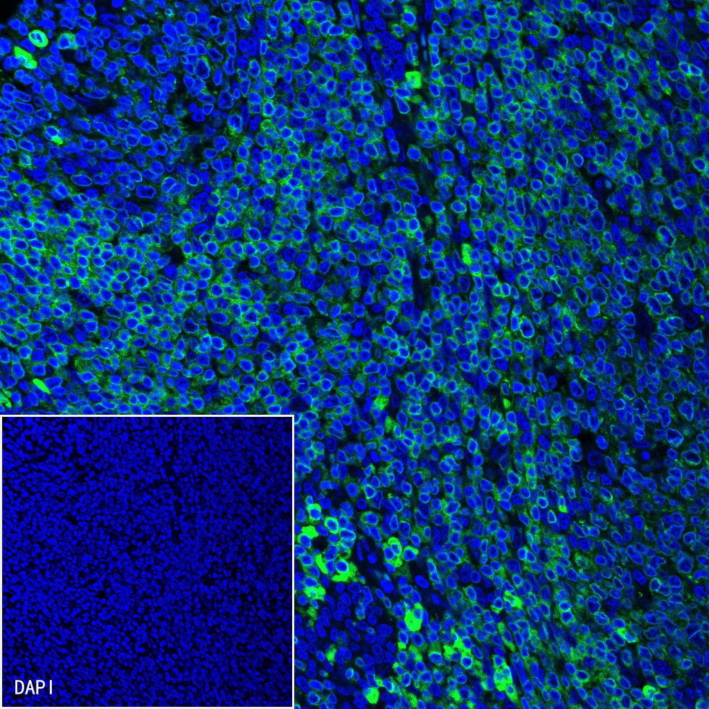
IF shows positive staining in paraffin-embedded human tonsil. Anti-CD79a/MB-1 antibody was used at 1/500 dilution (Green) and incubated overnight at 4°C. Goat polyclonal Antibody to Rabbit IgG - H&L (Alexa Fluor® 488) was used as secondary antibody at 1/1000 dilution. Counterstained with DAPI (Blue). Heat mediated antigen retrieval with EDTA buffer pH9.0 was performed before commencing with IF staining protocol.
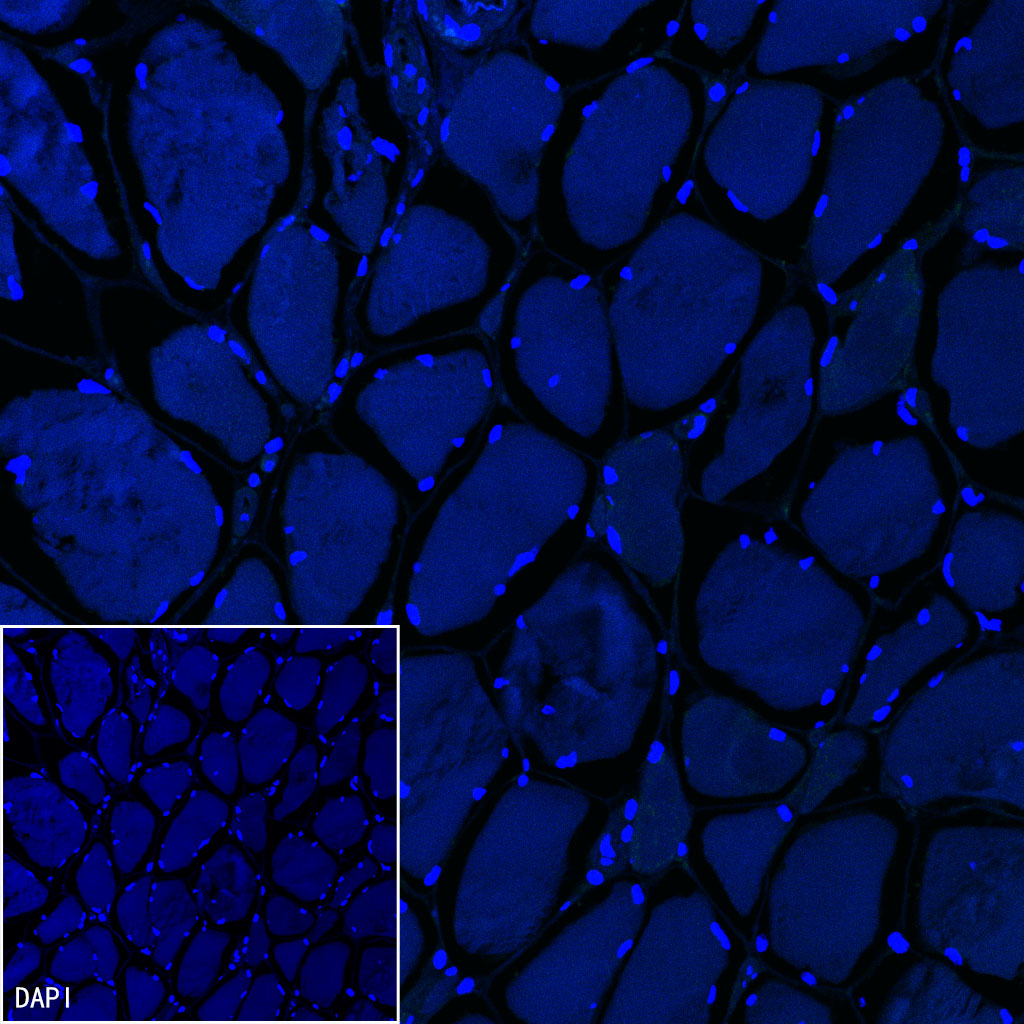
Negative control: IF shows negative staining in paraffin-embedded human skeletal muscle. Anti-CD79a/MB-1 antibody was used at 1/500 dilution and incubated overnight at 4°C. Goat polyclonal Antibody to Rabbit IgG - H&L (Alexa Fluor® 488) was used as secondary antibody at 1/1000 dilution. Counterstained with DAPI (Blue). Heat mediated antigen retrieval with EDTA buffer pH9.0 was performed before commencing with IF staining protocol.

评论(0)