


 申请试用
申请试用 产品介绍 评论(0)
宿主来源
Mouse抗原名称
CD68分子别名
KP1, PGM1, Macrosialin, Gp110免疫原
N/A细胞定位
Endosome, Membrane, LysosomeAccession
P34810克隆号
SDT-R146抗体类型
Recombinant mAb抗体同种型
IgG1反应种属 ?
Hu纯化方式
Protein G浓度
0.4 mg/ml标签
N/A性状
Liquid缓冲体系
PBS, 40% Glycerol, 0.05% BSA, 0.03% Proclin 300储存条件
12 months from date of receipt / reconstitution, -20 °C as supplied
应用
稀释度
应用 稀释度 IHC-P 1:1000 ICC 1:25
CD68 (Cluster of Differentiation 68) is a protein highly expressed by cells in the monocyte lineage (e.g., monocytic phagocytes, osteoclasts), by circulating macrophages, and by tissue macrophages. Immunohistochemistry can be used to identify the presence of CD68, which is found in the cytoplasmic granules of a range of different blood cells and myocytes. It is particularly useful as a marker for the various cells of the macrophage lineage, including monocytes, histiocytes, giant cells, Kupffer cells, and osteoclasts. This allows it to be used to distinguish diseases of otherwise similar appearance, such as the monocyte/macrophage and lymphoid forms of leukaemia (the latter being CD68 negative). Its presence in macrophages also makes it useful in diagnosing conditions related to proliferation or abnormality of these cells, such as malignant histiocytosis, histiocytic lymphoma, and Gaucher's disease.
免疫组化
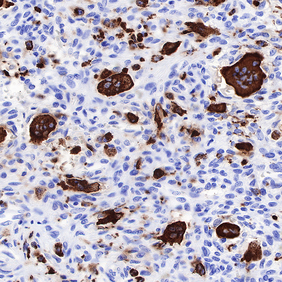
IHC shows positive staining in paraffin-embedded human giant cell tumor of bone. Anti-CD68 antibody was used at 1/1000 dilution, followed by a HRP Polymer for Mouse & Rabbit IgG (ready to use). Counterstained with hematoxylin. Heat mediated antigen retrieval with Tris/EDTA buffer pH9.0 was performed before commencing with IHC staining protocol.
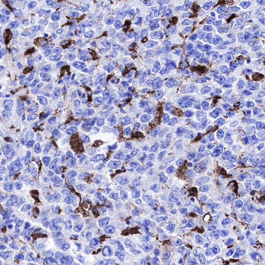
IHC shows positive staining in paraffin-embedded human diffuse large B-cell lymphoma. Anti-CD68 antibody was used at 1/1000 dilution, followed by a HRP Polymer for Mouse & Rabbit IgG (ready to use). Counterstained with hematoxylin. Heat mediated antigen retrieval with Tris/EDTA buffer pH9.0 was performed before commencing with IHC staining protocol.
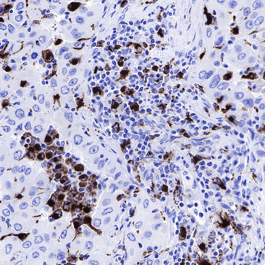
IHC shows positive staining in paraffin-embedded human lung adenocarcinoma. Anti-CD68 antibody was used at 1/1000 dilution, followed by a HRP Polymer for Mouse & Rabbit IgG (ready to use). Counterstained with hematoxylin. Heat mediated antigen retrieval with Tris/EDTA buffer pH9.0 was performed before commencing with IHC staining protocol.
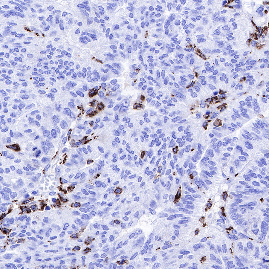
IHC shows positive staining in paraffin-embedded human ovarian carcinoma. Anti-CD68 antibody was used at 1/1000 dilution, followed by a HRP Polymer for Mouse & Rabbit IgG (ready to use). Counterstained with hematoxylin. Heat mediated antigen retrieval with Tris/EDTA buffer pH9.0 was performed before commencing with IHC staining protocol.
IHC shows positive staining in paraffin-embedded human tonsil. Anti-CD68 antibody was used at 1/1000 dilution, followed by a HRP Polymer for Mouse & Rabbit IgG (ready to use). Counterstained with hematoxylin. Heat mediated antigen retrieval with Tris/EDTA buffer pH9.0 was performed before commencing with IHC staining protocol.
IHC shows positive staining in paraffin-embedded human colon. Anti-CD68 antibody was used at 1/1000 dilution, followed by a HRP Polymer for Mouse & Rabbit IgG (ready to use). Counterstained with hematoxylin. Heat mediated antigen retrieval with Tris/EDTA buffer pH9.0 was performed before commencing with IHC staining protocol.
IHC shows positive staining in paraffin-embedded human lung. Anti-CD68 antibody was used at 1/1000 dilution, followed by a HRP Polymer for Mouse & Rabbit IgG (ready to use). Counterstained with hematoxylin. Heat mediated antigen retrieval with Tris/EDTA buffer pH9.0 was performed before commencing with IHC staining protocol.
Negative control: IHC shows negative staining in paraffin-embedded human gastrointestinal stromal tumor. Anti-CD68 antibody was used at 1/1000 dilution, followed by a HRP Polymer for Mouse & Rabbit IgG (ready to use). Counterstained with hematoxylin. Heat mediated antigen retrieval with Tris/EDTA buffer pH9.0 was performed before commencing with IHC staining protocol.
免疫细胞化学
ICC shows positive staining in RAW264.7 cells. Anti-CD68 antibody was used at 1/250 dilution (Green) and incubated overnight at 4°C. Goat polyclonal Antibody to Mouse IgG - H&L (Alexa Fluor® 488) was used as secondary antibody at 1/1000 dilution. The cells were fixed with 100% ice-cold methanol and permeabilized with 0.1% PBS-Triton X-100. Nuclei were counterstained with DAPI (Blue).

评论(0)