


 申请试用
申请试用 产品介绍 评论(0)
宿主来源
Mouse抗原名称
PD-1分子别名
Programmed cell death protein 1, hPD-1, CD279免疫原
N/A细胞定位
Cell membraneAccession
Q15116克隆号
SDT-R143抗体类型
Recombinant mAb抗体同种型
IgG1反应种属 ?
Hu纯化方式
Protein G浓度
2 mg/ml标签
N/A性状
Liquid缓冲体系
PBS, 40% Glycerol, 0.05% BSA, 0.03% Proclin 300
储存条件
12 months from date of receipt / reconstitution, -20 °C as supplied
应用
稀释度
应用 稀释度 IHC-P 1:200-1:500 ICFCM 1:200 ICC 1:100 WB 1:1000 IP 1:200 IF 1:200
Programmed cell death protein 1, also known as PD-1 and CD279 (cluster of differentiation 279), is a protein on the surface of T and B cells that has a role in regulating the immune system's response to the cells of the human body by down-regulating the immune system and promoting self-tolerance by suppressing T cell inflammatory activity. This prevents autoimmune diseases, but it can also prevent the immune system from killing cancer cells. PD-1 is an immune checkpoint and guards against autoimmunity through two mechanisms. First, it promotes apoptosis (programmed cell death) of antigen-specific T-cells in lymph nodes. Second, it reduces apoptosis in regulatory T cells (anti-inflammatory, suppressive T cells).
免疫印迹
WB result of PD-1 Mouse mAb
Primary antibody: PD-1 Mouse mAb at 1/1000 dilution
Lane 1: Jurkat whole cell lysate 20 µg
Lane 2: MOLT-4 whole cell lysate 20 µg
Lane 3: MOLT-4 treated with Ionomycin (500ng/ml 24 hr) and PMA (10ng/ml 24 hr) whole cell lysate 20 µg
Negative control: Jurkat whole cell lysate
Secondary antibody: Goat Anti-mouse IgG, (H+L), HRP conjugated at 1/10000 dilution
Predicted MW: 32 kDa
Observed MW: 38~70 kDa
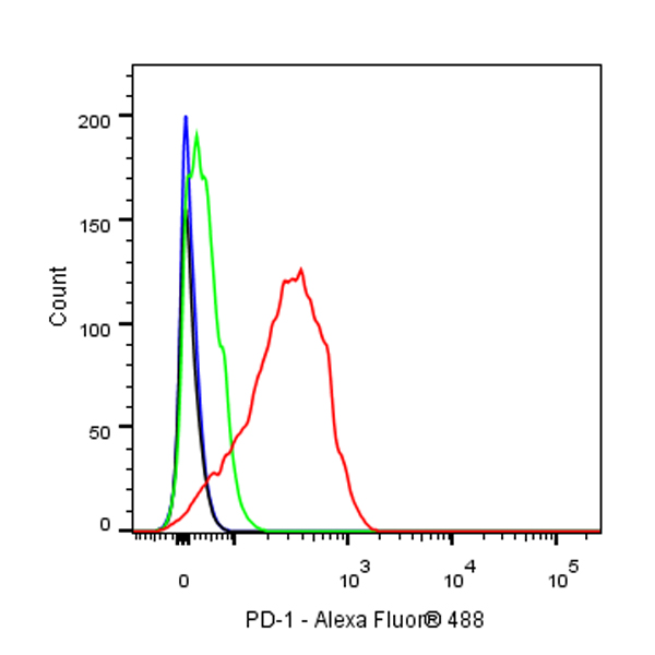
流式分析
Flow cytometric analysis of Molt-4 (Human cervix adenocarcinoma epithelial cell) cells, treated with 10ng/ml PMA and 500ng/ml Ionomycin for 24h (Red) or untreated (Green), labeling PD-1 at 1/200 dilution (1 μg) compared with a Mouse monoclonal IgG isotype control (Black) and an unlabeled control (cells without incubation with primary antibody and secondary antibody) (Blue). Goat Anti - Mouse IgG Alexa Fluor® 488 was used as the secondary antibody.
免疫沉淀
PD-1 Mouse mAb at 1/200 dilution (1 µg) immunoprecipitating PD-1 in 0.35 mg MOLT-4 treated with Ionomycin (500ng/ml 24 hr) and PMA (10ng/ml 24 hr) whole cell lysate.
Western blot was performed on the immunoprecipitate using PD-1 Mouse mAb at 1/1000 dilution.
Secondary antibody (HRP) for IP was used at 1/2000 dilution.
Lane 1: MOLT-4 treated with Ionomycin (500ng/ml 24 hr) and PMA (10ng/ml 24 hr) whole cell lysate 20 µg (Input)
Lane 2: PD-1 Mouse mAb IP in MOLT-4 treated with Ionomycin (500ng/ml 24 hr) and PMA (10ng/ml 24 hr) whole cell lysate
Lane 3: Mouse monoclonal IgG1 IP in MOLT-4 treated with Ionomycin (500ng/ml 24 hr) and PMA (10ng/ml 24 hr) whole cell lysate
Predicted MW: 32 kDa
Observed MW: 38~70 kDa
(This blot was developed with high sensitivity substrate)
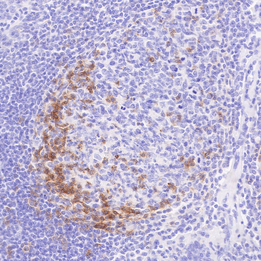
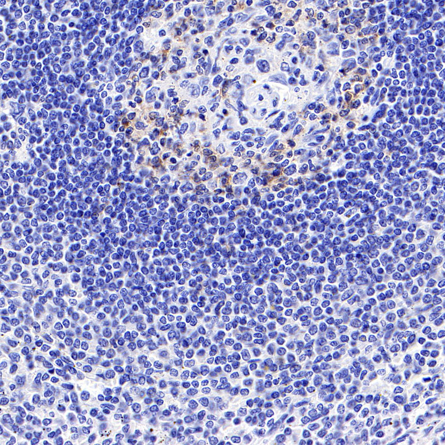
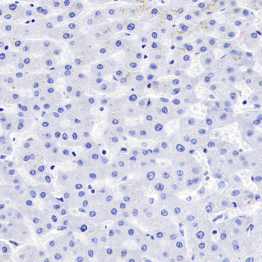
免疫组化
IHC shows positive staining in paraffin-embedded human tonsil. Anti-PD-1 antibody was used at 1/200 dilution, followed by a HRP Polymer for Mouse & Rabbit IgG (ready to use). Counterstained with hematoxylin. Heat mediated antigen retrieval with Tris/EDTA buffer pH9.0 was performed before commencing with IHC staining protocol.
IHC shows positive staining in paraffin-embedded human spleen. Anti-PD-1 antibody was used at 1/500 dilution, followed by a HRP Polymer for Mouse & Rabbit IgG (ready to use). Counterstained with hematoxylin. Heat mediated antigen retrieval with Tris/EDTA buffer pH9.0 was performed before commencing with IHC staining protocol.
Negative control: IHC shows negative staining in paraffin-embedded human liver. Anti-PD-1 antibody was used at 1/500 dilution, followed by a HRP Polymer for Mouse & Rabbit IgG (ready to use). Counterstained with hematoxylin. Heat mediated antigen retrieval with Tris/EDTA buffer pH9.0 was performed before commencing with IHC staining protocol.
免疫细胞化学
ICC shows positive staining in MOLT-4 cells treated with Ionomycin (500ng/ml,24h) + PMA (10ng/ml,24h). Anti-PD-1 antibody was used at 1/100 dilution (Green) and incubated overnight at 4°C. Goat polyclonal Antibody to mouse IgG - H&L (Alexa Fluor® 488) was used as secondary antibody at 1/1000 dilution. The cells were fixed with 4% PFA and permeabilized with 0.1% PBS-Triton X-100. Nuclei were counterstained with DAPI (Blue).
Negative control:ICC shows negative staining in MOLT-4 cells untreated with Ionomycin (500ng/ml,24h) + PMA (10ng/ml,24h). Anti-PD-1 antibody was used at 1/100 dilution and incubated overnight at 4°C. Goat polyclonal Antibody to mouse IgG - H&L (Alexa Fluor® 488) was used as secondary antibody at 1/1000 dilution. The cells were fixed with 4% PFA and permeabilized with 0.1% PBS-Triton X-100. Nuclei were counterstained with DAPI (Blue).
免疫荧光
IF shows positive staining in paraffin-embedded human tonsil. Anti-PD-1 antibody was used at 1/200 dilution (magenta) and incubated overnight at 4°C. Goat polyclonal Antibody to Mouse IgG - H&L (Alexa Fluor®647) was used as secondary antibody at 1/500 dilution. Counterstained with DAPI (Blue). Heat mediated antigen retrieval with EDTA buffer pH9.0 was performed before commencing with IF staining protocol.
Negative control: IF shows negative staining in paraffin-embedded human liver. Anti-PD-1 antibody was used at 1/200 dilution and incubated overnight at 4°C. Goat polyclonal Antibody to Mouse IgG - H&L (Alexa Fluor®647) was used as secondary antibody at 1/500 dilution. Counterstained with DAPI (Blue). Heat mediated antigen retrieval with EDTA buffer pH9.0 was performed before commencing with IF staining protocol.

评论(0)