


 申请试用
申请试用 产品介绍 引用文献(2) 评论(0)
宿主来源
Rabbit抗原名称
CD4分子别名
T-cell surface glycoprotein CD4, T-cell surface antigen T4/Leu-3, OKT4免疫原
N/A细胞定位
Cell membraneAccession
P01730克隆号
SDT-R163抗体类型
Recombinant mAb反应种属 ?
Hu纯化方式
Protein A浓度
0.5 mg/ml性状
Liquid缓冲体系
PBS, 40% Glycerol, 0.05% BSA, 0.03% Proclin 300储存条件
12 months from date of receipt / reconstitution, -20 °C as supplied
应用
IHC-P ? ,FCM ,WB ,IP ,IF ?稀释度
应用 稀释度 WB 1:1000 IHC-P 1:500 FCM 1:50 IP 1:50 IF 1:200
CD4 (cluster of differentiation 4) is a glycoprotein found on the surface of immune cells such as T helper cells, monocytes, macrophages, and dendritic cells. It is a type of white blood cell that helps fight infection by triggering your immune system to destroy viruses, bacteria, and other germs that may make you sick. CD4 is also a receptor for the HIV virus, and when the virus infects cells with CD4 surface proteins, it depletes the number of T cells, B cells, natural killer cells, and monocytes in the patient's blood.
免疫印迹
- WB result of CD4 Rabbit mAb Primary antibody: CD4 Rabbit mAb at 1/1000 dilution Lane 1: Ramos whole cell lysate 20 µg Lane 2: THP-1 whole cell lysate 20 µg Lane 3: Jurkat whole cell lysate 20 µg Lane 4: Molt-4 whole cell lysate 20 µg Negative control: Ramos whole cell lysate Secondary antibody: Goat Anti-Rabbit IgG, (H+L), HRP conjugated at 1/10000 dilution Predicted MW: 51kDa Observed MW: 55kDa
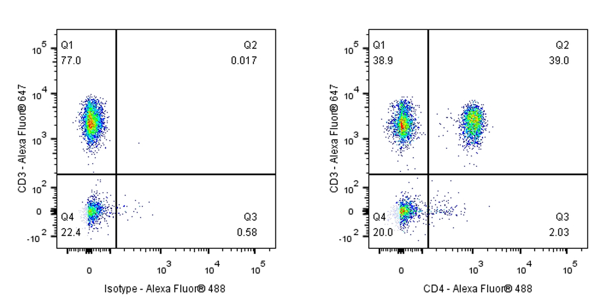
流式分析
Flow cytometric analysis of human PBMC (human peripheral blood mononuclear cell) labelling CD4 antibody at 1/50 (1 μg) dilution (Right) compared with a Rabbit monoclonal IgG isotype control (Left). Goat Anti – Rabbit IgG Alexa Fluor® 488 was used as the secondary antibody, cells were stained with CD3 - Alexa Fluor® 647 simultaneously. Events were gated on viable lymphocytes.
免疫沉淀
CD4 Rabbit mAb at 1/50 dilution (1 µg) immunoprecipitating CD4 in 0.4 mg THP-1 whole cell lysate.
Western blot was performed on the immunoprecipitate using CD4 Rabbit mAb at 1/1000 dilution.
Secondary antibody (HRP) for IP was used at 1/400 dilution.
Lane 1: THP-1 whole cell lysate 20 µg (Input)
Lane 2: CD4 Rabbit mAb IP in THP-1 whole cell lysate
Lane 3: Rabbit monoclonal IgG IP in THP-1 whole cell lysate
Predicted MW: 51 kDa
Observed MW: 55 kDa
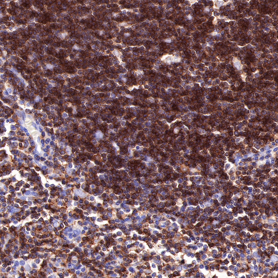
免疫组化
- IHC shows positive staining in paraffin-embedded human thymus. Anti-CD4 antibody was used at 1/500 dilution, followed by a HRP Polymer for Mouse & Rabbit IgG (ready to use). Counterstained with hematoxylin. Heat mediated antigen retrieval with Tris/EDTA buffer pH9.0 was performed before commencing with IHC staining protocol.
- IHC shows positive staining in paraffin-embedded human tonsil. Anti-CD4 antibody was used at 1/500 dilution, followed by a HRP Polymer for Mouse & Rabbit IgG (ready to use). Counterstained with hematoxylin. Heat mediated antigen retrieval with Tris/EDTA buffer pH9.0 was performed before commencing with IHC staining protocol.
- IHC shows positive staining in paraffin-embedded human spleen. Anti-CD4 antibody was used at 1/500 dilution, followed by a HRP Polymer for Mouse & Rabbit IgG (ready to use). Counterstained with hematoxylin. Heat mediated antigen retrieval with Tris/EDTA buffer pH9.0 was performed before commencing with IHC staining protocol.
- IHC shows positive staining in paraffin-embedded human liver. Anti-CD4 antibody was used at 1/500 dilution, followed by a HRP Polymer for Mouse & Rabbit IgG (ready to use). Counterstained with hematoxylin. Heat mediated antigen retrieval with Tris/EDTA buffer pH9.0 was performed before commencing with IHC staining protocol.
- IHC shows positive staining in paraffin-embedded human cervical squamous cell carcinoma. Anti-CD4 antibody was used at 1/500 dilution, followed by a HRP Polymer for Mouse & Rabbit IgG (ready to use). Counterstained with hematoxylin. Heat mediated antigen retrieval with Tris/EDTA buffer pH9.0 was performed before commencing with IHC staining protocol.
- Negative control: IHC shows negative staining in paraffin-embedded human cerebral cortex. Anti-CD4 antibody was used at 1/500 dilution, followed by a HRP Polymer for Mouse & Rabbit IgG (ready to use). Counterstained with hematoxylin. Heat mediated antigen retrieval with Tris/EDTA buffer pH9.0 was performed before commencing with IHC staining protocol.
免疫荧光
IF shows positive staining in paraffin-embedded human tonsil. Anti-CD4 antibody was used at 1/200 dilution (Green) and incubated overnight at 4°C. Goat polyclonal Antibody to Rabbit IgG - H&L (Alexa Fluor® 488) was used as secondary antibody at 1/1000 dilution. Counterstained with DAPI (Blue). Heat mediated antigen retrieval with EDTA buffer pH9.0 was performed before commencing with IF staining protocol.
引用文献(2)
- Integrated analysis identifies CD276 in fibroblasts as a malignancy predictor and regulator of neutrophil infiltration in hepatoblastoma
Miao Ding,Guoqing Zhu,Tianshu Chen,Jiabei Zhu,Siwei Mao,Xiaochen Tang,Han Wu,Ni Zhen,Fenyong Sun,Qiuhui Pan,Ji Ma
Hepatol Commun. 2025 Oct 31 ; 41171428
影响因子: 4.6
货号:S0B2179产品名称:CD4 Recombinant Rabbit mAb (SDT-R163)
- Neoadjuvant PD-1 blockade induces the autophagy of immune cells: a new target for synergistic therapy of recurrent glioblastoma
Z Xuan, K Wang, Q Zhu, T Sun, J Jiang, Z Wu
Biochemistry and Biophysics Reports. 2025 Sep 01 .
影响因子: 2.2
货号:S0B2179产品名称:CD4 Recombinant Rabbit mAb (SDT-R163)

评论(0)