


 大包装询价
大包装询价 产品介绍 评论(0)
宿主来源
Rabbit抗原名称
Myosin-11/SMMHC分子别名
smooth muscle myosin heavy chain, HSM-V, SMMS-1, SM-MYOSIN-H, Myosin heavy chain 11, MYH11免疫原
Synthetic Peptide细胞定位
MelanosomeAccession
P35749克隆号
SDT-188-112抗体类型
Recombinant mAb应用
IHC-P, WB, IF反应种属 ?
Hu, Ms, Rt预测反应种属
(反应种属缩写表)Rb纯化方式
Protein A浓度
0.25 mg/ml性状
Liquid缓冲体系
PBS, 40% Glycerol, 0.05% BSA, 0.03% Proclin 300储存条件
12 months from date of receipt / reconstitution, -20 °C as supplied
| 应用 | 稀释度 |
|---|---|
| WB | 1:1000-1:25000 |
| IHC-P | 1:1000 |
| IF | 1:125 |
SMMHC is a structural component of the smooth muscle myosin molecule and is a specific marker of “terminal” smooth muscle differentiation [PMID: 10721417, PMID: 3882826]. SMMHC is composed of at least two isoforms: SM1 (204 kDa) and SM2 (200 kDa), both of which are encoded by a single gene [PMID: 9702855, PMID: 10998642]. The SM1 isoform is expressed in the MEC of normal mammary glands, fibrocystic diseases, and in myoepithelial derived tumours of the breast [PMID: 11493962, PMID: 9702855]. Furthermore, studies have documented that antibodies to SMMHC and calponin, both markers of terminal smooth muscle differentiation, are more specific for breast MEC than are other more commonly used antibodies, such as those that recognise SMA [PMID: 11493962].
免疫印迹
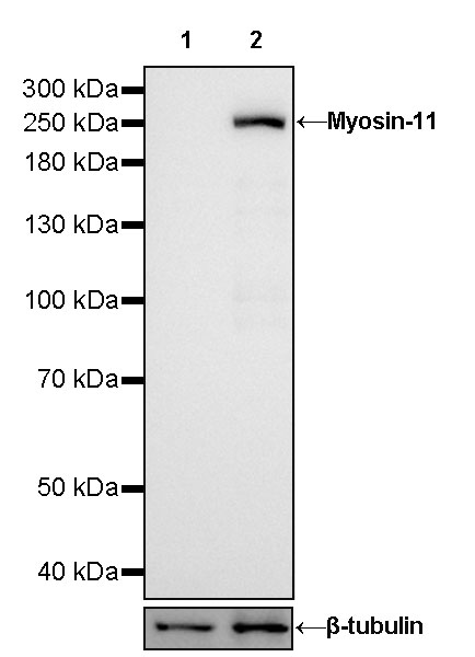 WB result of Myosin-11 Rabbit mAb Primary antibody: Myosin-11 Rabbit mAb at 1/25000 dilution Lane 1: mouse skeletal muscle lysate 5 µg Lane 2: mouse bladder lysate 5 µg Negative control: mouse skeletal muscle lysate Secondary antibody: Goat Anti-Rabbit IgG, (H+L), HRP conjugated at 1/10000 dilution Predicted MW: 227kDa Observed MW: 250kDa Exposure time: 30s
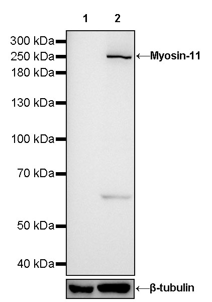 WB result of Myosin-11 Rabbit mAb Primary antibody: Myosin-11 Rabbit mAb at 1/1000 dilution Lane 1: mouse skeletal muscle lysate 20 µg Lane 2: mouse prostate lysate 20 µg Negative control: mouse skeletal muscle lysate Secondary antibody: Goat Anti-Rabbit IgG, (H+L), HRP conjugated at 1/10000 dilution Predicted MW: 227kDa Observed MW: 250kDa Exposure time: 60s
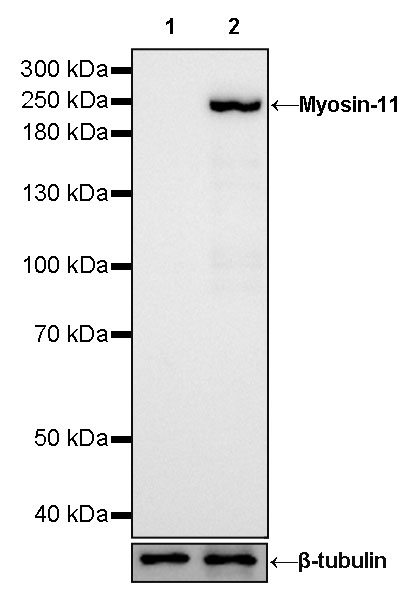 WB result of Myosin-11 Rabbit mAb Primary antibody: Myosin-11 Rabbit mAb at 1/25000 dilution Lane 1: rat skeletal muscle lysate 5 µg Lane 2: rat bladder lysate 5 µg Negative control: rat skeletal muscle lysate Secondary antibody: Goat Anti-Rabbit IgG, (H+L), HRP conjugated at 1/10000 dilution Predicted MW: 227kDa Observed MW: 250kDa Exposure time: 30s
免疫组化
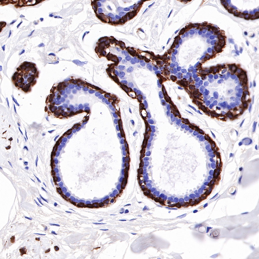 IHC shows positive staining in paraffin-embedded human breast. Anti-Myosin-11 antibody was used at 1/1000 dilution, followed by a HRP Polymer for Mouse & Rabbit IgG (ready to use). Counterstained with hematoxylin. Heat mediated antigen retrieval with Tris/EDTA buffer pH9.0 was performed before commencing with IHC staining protocol.
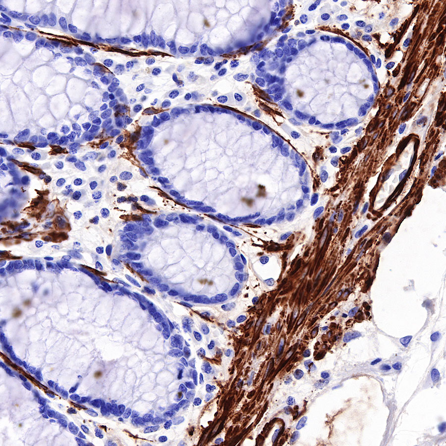 IHC shows positive staining in paraffin-embedded human colon. Anti-Myosin-11 antibody was used at 1/1000 dilution, followed by a HRP Polymer for Mouse & Rabbit IgG (ready to use). Counterstained with hematoxylin. Heat mediated antigen retrieval with Tris/EDTA buffer pH9.0 was performed before commencing with IHC staining protocol.
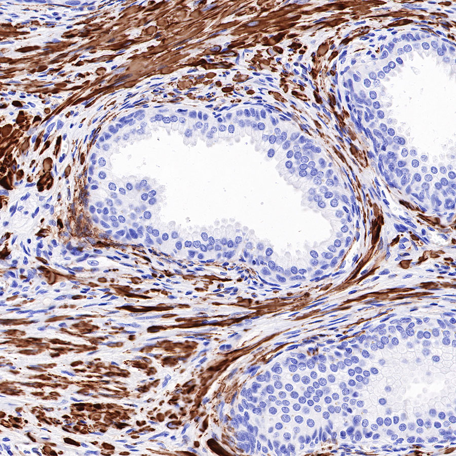 IHC shows positive staining in paraffin-embedded human prostate. Anti-Myosin-11 antibody was used at 1/1000 dilution, followed by a HRP Polymer for Mouse & Rabbit IgG (ready to use). Counterstained with hematoxylin. Heat mediated antigen retrieval with Tris/EDTA buffer pH9.0 was performed before commencing with IHC staining protocol.
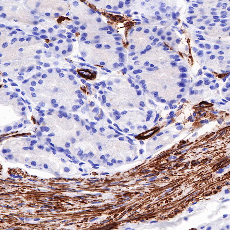 IHC shows positive staining in paraffin-embedded human stomach. Anti-Myosin-11 antibody was used at 1/1000 dilution, followed by a HRP Polymer for Mouse & Rabbit IgG (ready to use). Counterstained with hematoxylin. Heat mediated antigen retrieval with Tris/EDTA buffer pH9.0 was performed before commencing with IHC staining protocol.
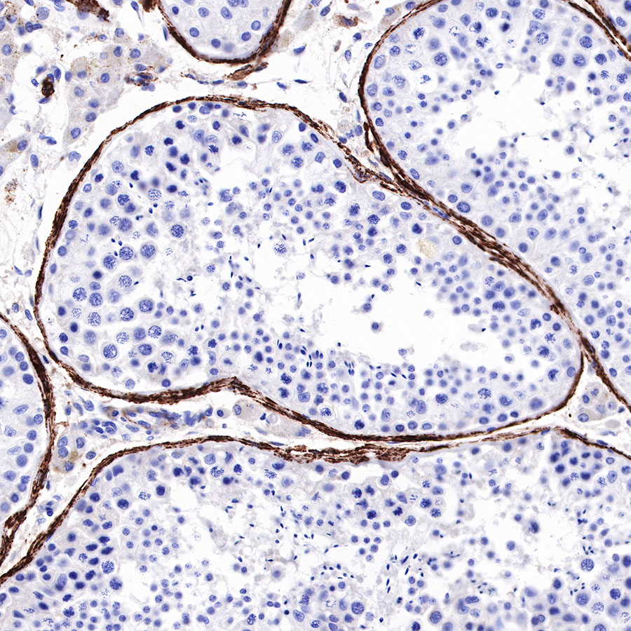 IHC shows positive staining in paraffin-embedded human testis. Anti-Myosin-11 antibody was used at 1/1000 dilution, followed by a HRP Polymer for Mouse & Rabbit IgG (ready to use). Counterstained with hematoxylin. Heat mediated antigen retrieval with Tris/EDTA buffer pH9.0 was performed before commencing with IHC staining protocol.
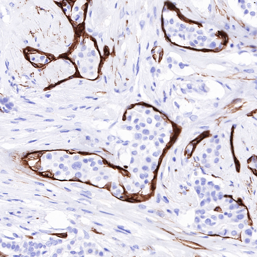 IHC shows positive staining in paraffin-embedded human breast cancer. Anti-Myosin-11 antibody was used at 1/1000 dilution, followed by a HRP Polymer for Mouse & Rabbit IgG (ready to use). Counterstained with hematoxylin. Heat mediated antigen retrieval with Tris/EDTA buffer pH9.0 was performed before commencing with IHC staining protocol.
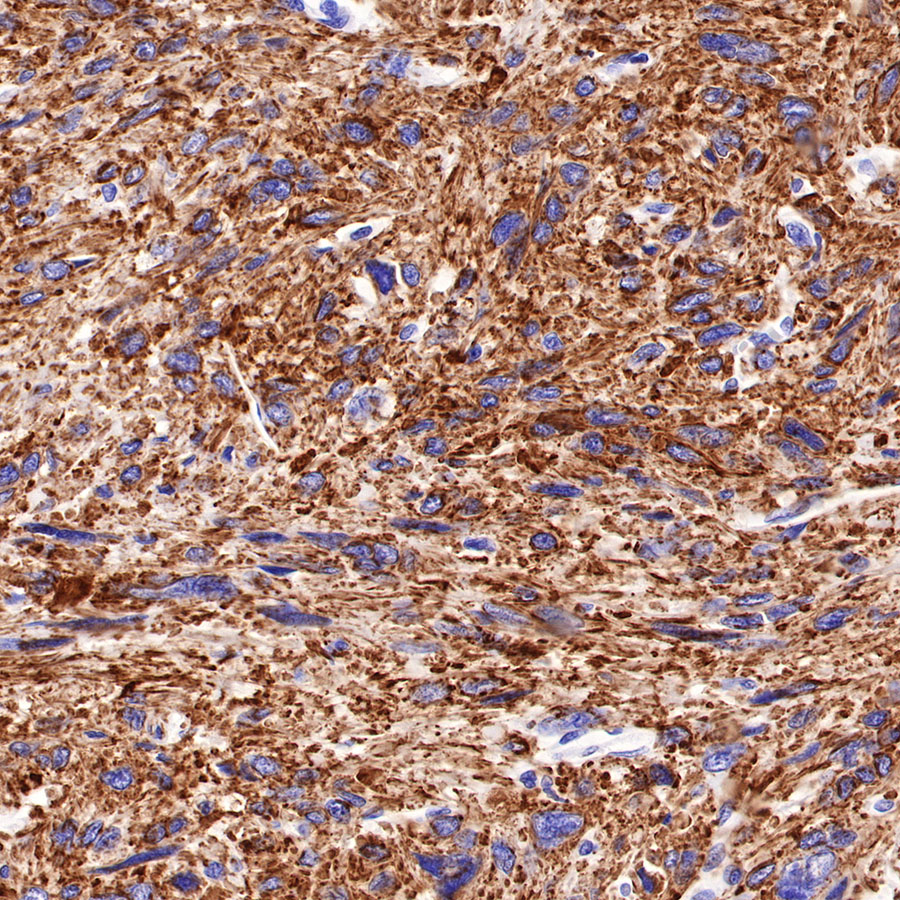 IHC shows positive staining in paraffin-embedded human leiomyosarcoma. Anti-Myosin-11 antibody was used at 1/1000 dilution, followed by a HRP Polymer for Mouse & Rabbit IgG (ready to use). Counterstained with hematoxylin. Heat mediated antigen retrieval with Tris/EDTA buffer pH9.0 was performed before commencing with IHC staining protocol.
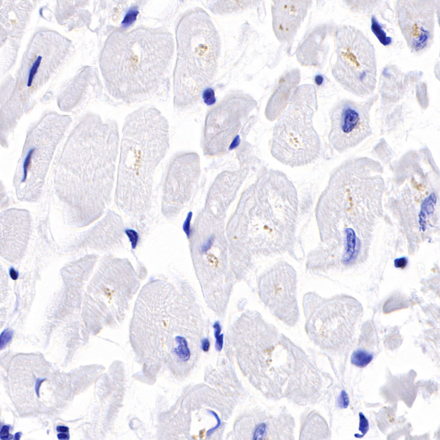 Negative control: IHC shows negative staining in paraffin-embedded human cardiac muscle. Anti-Myosin-11 antibody was used at 1/1000 dilution, followed by a HRP Polymer for Mouse & Rabbit IgG (ready to use). Counterstained with hematoxylin. Heat mediated antigen retrieval with Tris/EDTA buffer pH9.0 was performed before commencing with IHC staining protocol.
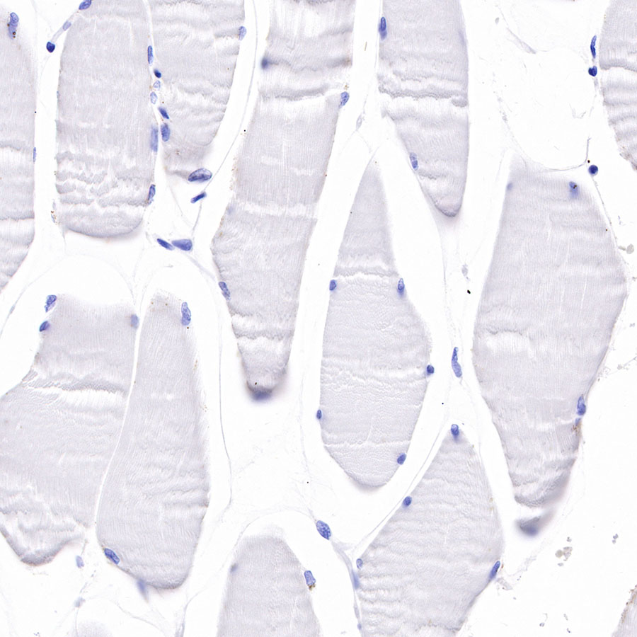 Negative control: IHC shows negative staining in paraffin-embedded human skeletal muscle. Anti-Myosin-11 antibody was used at 1/1000 dilution, followed by a HRP Polymer for Mouse & Rabbit IgG (ready to use). Counterstained with hematoxylin. Heat mediated antigen retrieval with Tris/EDTA buffer pH9.0 was performed before commencing with IHC staining protocol.
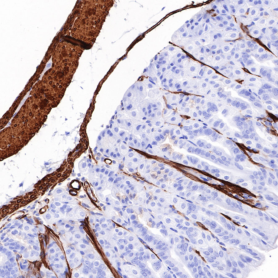 IHC shows positive staining in paraffin-embedded mouse stomach. Anti-Myosin-11 antibody was used at 1/1000 dilution, followed by a HRP Polymer for Mouse & Rabbit IgG (ready to use). Counterstained with hematoxylin. Heat mediated antigen retrieval with Tris/EDTA buffer pH9.0 was performed before commencing with IHC staining protocol.
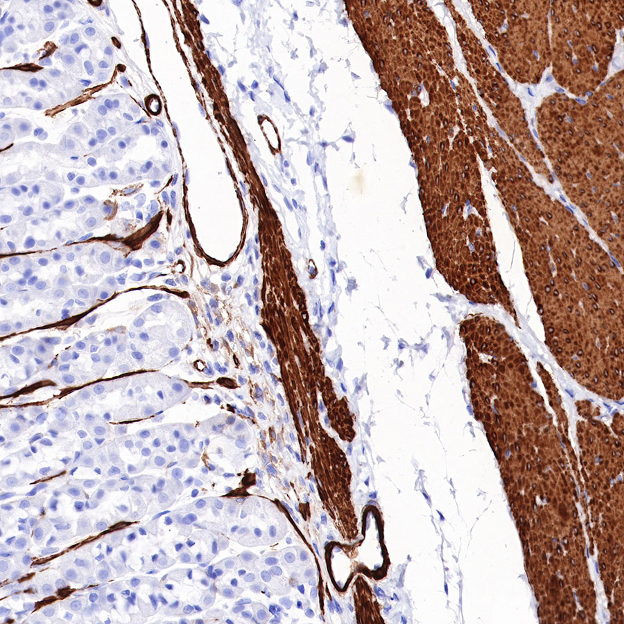 IHC shows positive staining in paraffin-embedded rat stomach. Anti-Myosin-11 antibody was used at 1/1000 dilution, followed by a HRP Polymer for Mouse & Rabbit IgG (ready to use). Counterstained with hematoxylin. Heat mediated antigen retrieval with Tris/EDTA buffer pH9.0 was performed before commencing with IHC staining protocol.
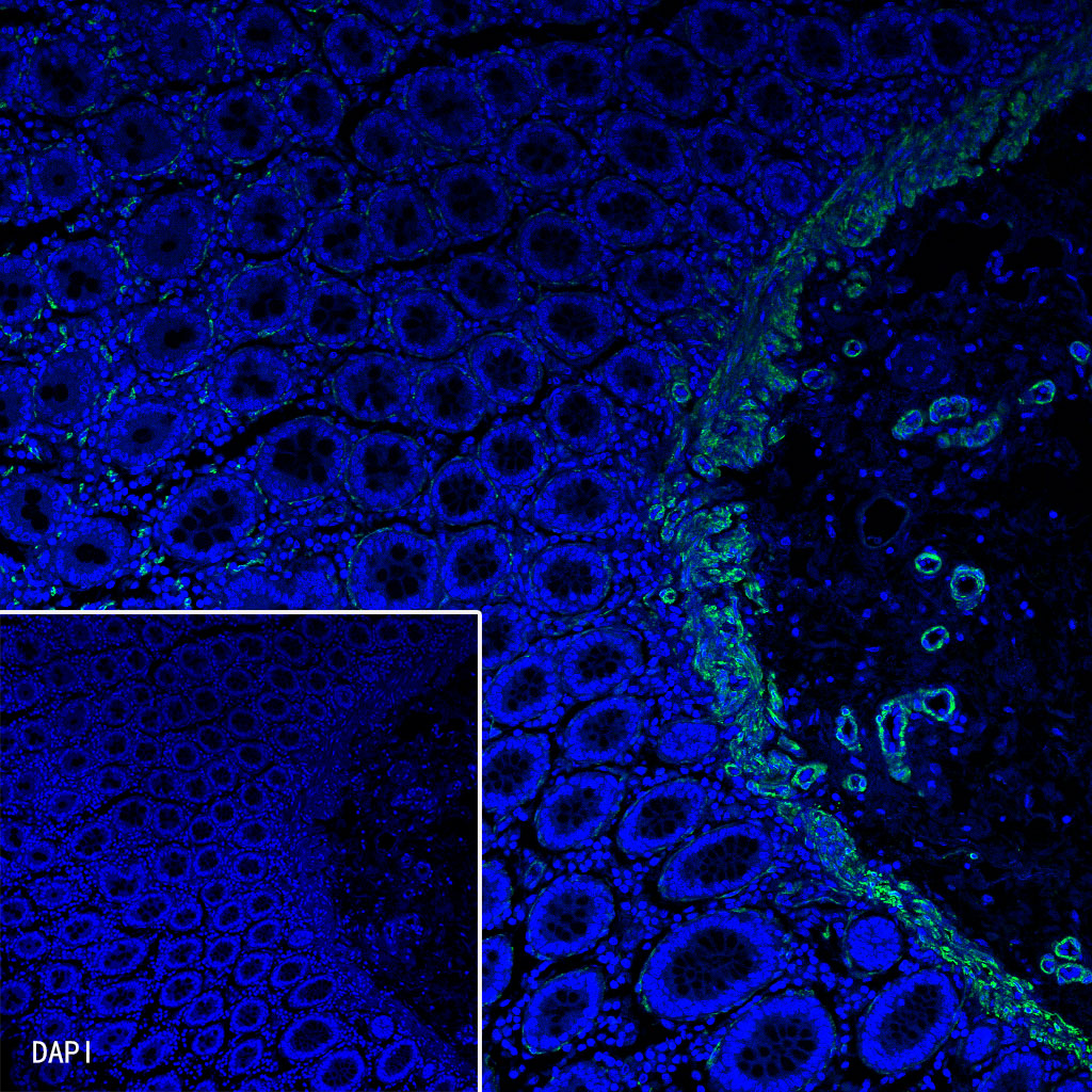 IF shows positive staining in paraffin-embedded human colon. Anti-Myosin-11 antibody was used at 1/125 dilution (Green). Goat polyclonal Antibody to Rabbit IgG - H&L (Alexa Fluor® 488) was used as secondary antibody at 1/1000 dilution. Counterstained with DAPI (Blue). Heat mediated antigen retrieval with Tris/EDTA buffer pH9.0 was performed before commencing with IF staining protocol.
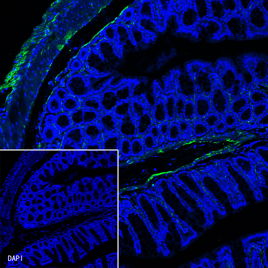 IF shows positive staining in paraffin-embedded mouse colon. Anti-Myosin-11 antibody was used at 1/125 dilution (Green). Goat polyclonal Antibody to Rabbit IgG - H&L (Alexa Fluor® 488) was used as secondary antibody at 1/1000 dilution. Counterstained with DAPI (Blue). Heat mediated antigen retrieval with Tris/EDTA buffer pH9.0 was performed before commencing with IF staining protocol.

评论(0)