




 分享
分享产品介绍 评论(0)
宿主来源
Rabbit抗原名称
LAG3分子别名
Lymphocyte activation gene 3 protein细胞定位
Cell membraneAccession
P18627克隆号
SDT-R159抗体类型
Recombinant mAb反应种属 ?
Hu纯化方式
Protein A浓度
1 mg/ml性状
Liquid缓冲体系
PBS
储存条件
12 months from date of receipt, 4°C as supplied
应用
IHC-P ?
IF ?
稀释度
应用 稀释度 IHC-P 1:1000 IF 1:500
LAG3 (CD223) is the third IR to be targeted in the clinic. It is a potential cancer immunotherapeutic target due to its negative regulatory role on T cells and its capacity, in combination with PD1, to mediate a state of exhaustion [PMID: 26018646]. LAG3 up-regulation is required to control overt activation and prevent the onset of autoimmunity. However, persistent antigen exposure in the tumor microenvironment results in sustained LAG3 expression, contributing to a state of exhaustion manifest in impaired proliferation and cytokine production. The exact signaling mechanisms downstream of LAG3 and interplay with other IRs remains largely unknown. However, the striking synergy between LAG3 and PD1 observed in multiple settings, coupled with the contrasting intracellular cytoplasmic domain of LAG3 as compared with other IRs, highlights the potential uniqueness of LAG3. There are now four LAG3-targeted therapies in the clinic with many more in preclinical development [PMID: 28258692].
免疫组化
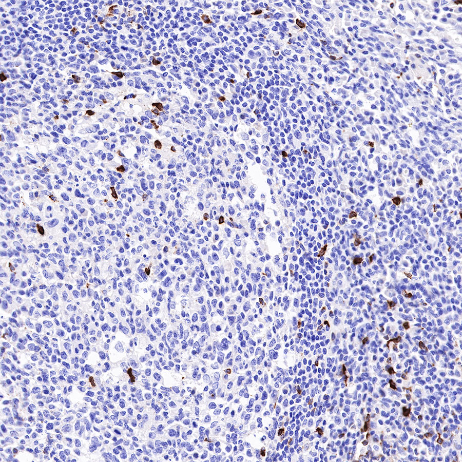
- IHC shows positive staining in paraffin-embedded human tonsil. Anti-LAG3 antibody was used at 1/1000 dilution, followed by a HRP Polymer for Mouse & Rabbit IgG (ready to use). Counterstained with hematoxylin. Heat mediated antigen retrieval with Tris/EDTA buffer pH9.0 was performed before commencing with IHC staining protocol.
- IHC shows positive staining in paraffin-embedded human Hodgkin's lymphoma. Anti-LAG3 antibody was used at 1/1000 dilution, followed by a HRP Polymer for Mouse & Rabbit IgG (ready to use). Counterstained with hematoxylin. Heat mediated antigen retrieval with Tris/EDTA buffer pH9.0 was performed before commencing with IHC staining protocol.
- IHC shows positive staining in paraffin-embedded human tonsil. Anti-LAG3 antibody was used at 1/1000 dilution, followed by a HRP Polymer for Mouse & Rabbit IgG (ready to use). Counterstained with hematoxylin. Heat mediated antigen retrieval with Tris/EDTA buffer pH9.0 was performed before commencing with IHC staining protocol.
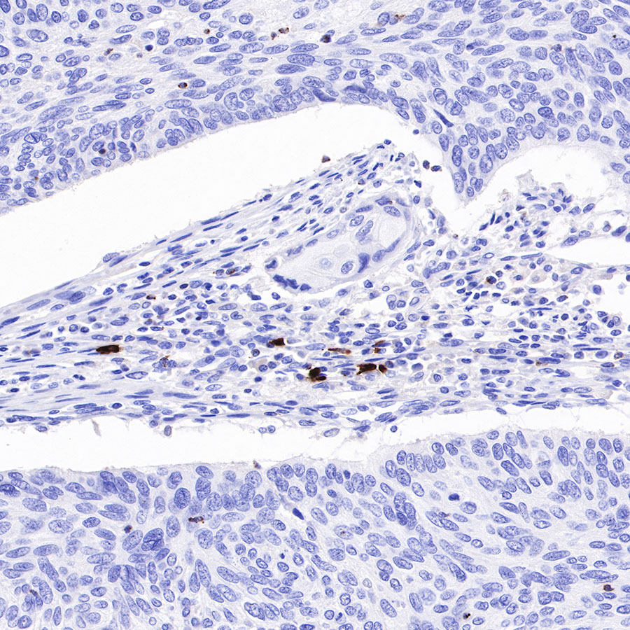
- IHC shows positive staining in paraffin-embedded human bladder cancer. Anti-LAG3 antibody was used at 1/1000 dilution, followed by a HRP Polymer for Mouse & Rabbit IgG (ready to use). Counterstained with hematoxylin. Heat mediated antigen retrieval with Tris/EDTA buffer pH9.0 was performed before commencing with IHC staining protocol.
- IHC shows positive staining in paraffin-embedded human thyroid cancer. Anti-LAG3 antibody was used at 1/1000 dilution, followed by a HRP Polymer for Mouse & Rabbit IgG (ready to use). Counterstained with hematoxylin. Heat mediated antigen retrieval with Tris/EDTA buffer pH9.0 was performed before commencing with IHC staining protocol.
免疫荧光
Flow cytometric analysis of Human CD326 expression on 4% PFA fixed 90% methanol permeabilized HeLa cells. Cells from the Hela (Human cervix adenocarcinoma epithelial cells) were stained with either Cy5 Rabbit IgG Isotype Control (Black line histogram) or SDT β-actin Recombinant Rabbit mAb (Cy5 Conjugate) (Red line histogram) at 0.1 μg/test, cells without incubation with primary antibody and secondary antibody (Blue line histogram) was used as unlabelled control. Flow cytometry and data analysis were performed using BD FACSymphony™ A1 and FlowJo™ software.
Flow cytometric analysis of Human CD326 expression on 4% PFA fixed 90% methanol permeabilized NIH/3T3 cells. Cells from the NIH/3T3 (Mouse embryonic fibroblast) were stained with either Cy5 Rabbit IgG Isotype Control (Black line histogram) or SDT β-actin Recombinant Rabbit mAb (Cy5 Conjugate) (Red line histogram) at 0.1 μg/test, cells without incubation with primary antibody and secondary antibody (Blue line histogram) was used as unlabelled control. Flow cytometry and data analysis were performed using BD FACSymphony™ A1 and FlowJo™ software.

评论(0)