


 申请试用
申请试用 产品介绍 评论(0)
宿主来源
Rabbit抗原名称
c-Myc分子别名
N-MYC, MYC, Transcription factor p64, bHLHe39免疫原
N/A细胞定位
NucleusAccession
P01106克隆号
SDT-R138反应种属 ?
Hu, Ms, Rt纯化方式
Protein A浓度
0.5 mg/ml性状
Liquid缓冲体系
PBS, 40% Glycerol, 0.05% BSA, 0.03% Proclin 300
储存条件
12 months from date of receipt / reconstitution, -20 °C as supplied
应用
稀释度
应用 稀释度 推荐种属 WB 1:1000 Hu, Ms IHC-P 1:500-1:1000 Hu, Ms, Rt ICFCM 1:50 Hu ICC 1:500 Hu ChIP 1:20-1:50 Hu
c-Myc (MYC) is a helix-loop-helix leucine zipper transcription factor that dimerizes with its partner protein Max to bind specific DNA sequences and transactivates genes. The Myc-Max heterodimer can also repress gene expression through complex formation with the transcription factor Miz1 [PubMed: 18923074]. In addition to its role in cancer, Myc is one of four transcription factors that collectively can re-program differentiated adult cells back to a pluripotent stem cell state [PubMed: 16904174]. Myc also plays an important role in normal cell physiology. The key distinction between physiological and oncogenic Myc function is whether MYC expression is regulated by normal circuitries, such as growth factor signaling that occurs when cells enter into the cell cycle and proliferate for tissue repair or whether, as in cancers, MYC activation can be short-circuited by genetic alterations, permitting deregulated Myc expression to alter transcription that no longer responds to external cues, particularly negative regulatory ones [PubMed: 16267388].
免疫印迹
WB result of c-Myc Rabbit mAb
Primary antibody: c-Myc Rabbit mAb at 1/1000 dilution
Lane 1: Neuro-2a whole cell lysate 20 µg
Secondary antibody: Goat Anti-Rabbit IgG, (H+L), HRP conjugated at 1/10000 dilution
Predicted MW: 49 kDa
Observed MW: 60 kDa
WB result of c-Myc Rabbit mAb
Primary antibody: c-Myc Rabbit mAb at 1/1000 dilution
Lane 1: HeLa whole cell lysate 20 µg
Lane 2: Jurkat whole cell lysate 20 µg
Secondary antibody: Goat Anti-Rabbit IgG, (H+L), HRP conjugated at 1/10000 dilution
Predicted MW: 49 kDa
Observed MW: 45, 57 kDa
流式分析
Flow cytometric analysis of 4% PFA fixed 90% methanol permeabilized HeLa (Human cervix adenocarcinoma epithelial cell) labelling c-Myc antibody at 1/50 dilution (1 μg)/ (Right) compared with a Rabbit monoclonal IgG / (Left) isotype control. Goat Anti - Rabbit IgG Alexa Fluor® 488 was used as the secondary antibody.
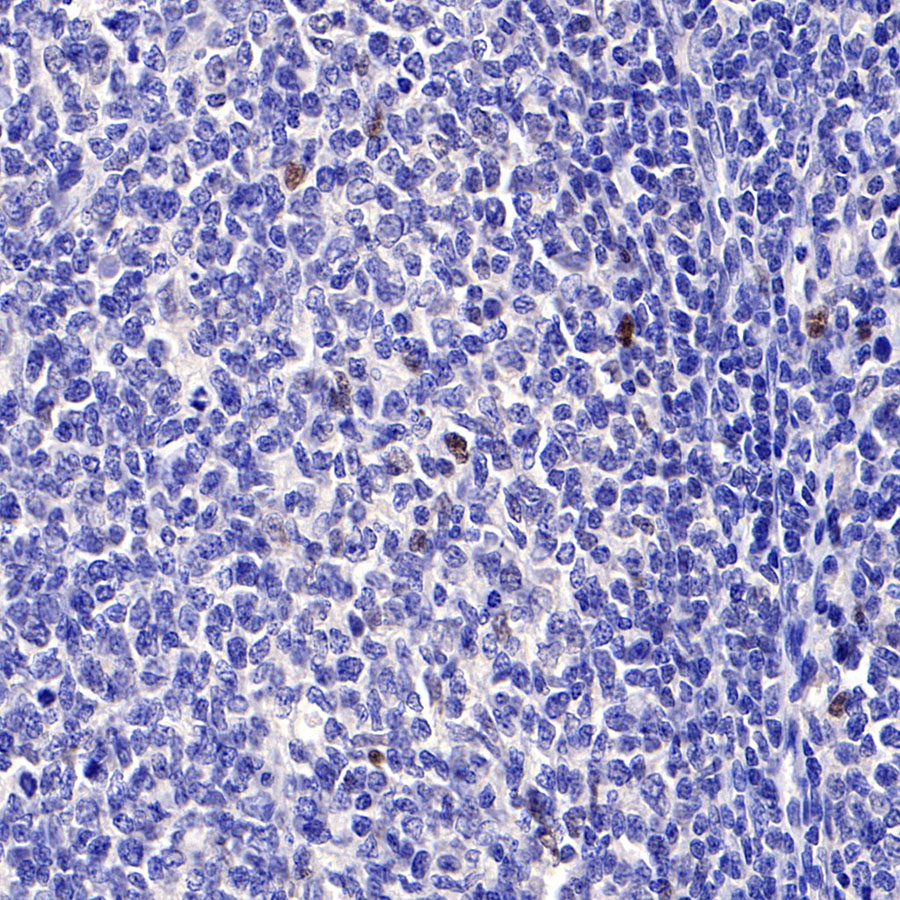
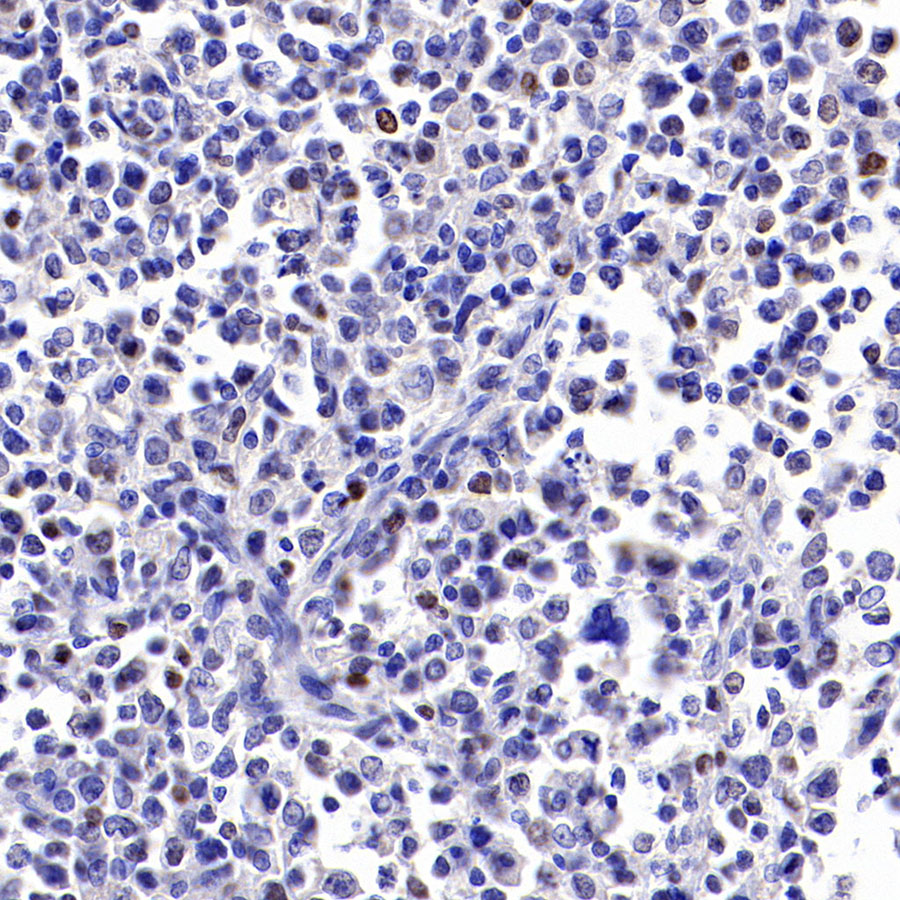
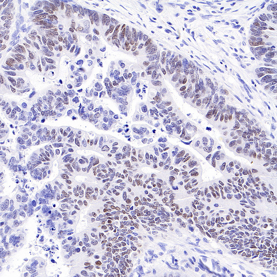
免疫组化
- IHC shows positive staining in paraffin-embedded human tonsil. Anti-c-Myc antibody was used at 1/500 dilution, followed by a HRP Polymer for Mouse & Rabbit IgG (ready to use). Counterstained with hematoxylin. Heat mediated antigen retrieval with Tris/EDTA buffer pH9.0 was performed before commencing with IHC staining protocol.
- IHC shows positive staining in paraffin-embedded human diffuse large B-cell lymphoma. Anti-c-Myc antibody was used at 1/500 dilution, followed by a HRP Polymer for Mouse & Rabbit IgG (ready to use). Counterstained with hematoxylin. Heat mediated antigen retrieval with Tris/EDTA buffer pH9.0 was performed before commencing with IHC staining protocol.
- IHC shows positive staining in paraffin-embedded human Hodgkin's lymphoma. Anti-c-Myc antibody was used at 1/500 dilution, followed by a HRP Polymer for Mouse & Rabbit IgG (ready to use). Counterstained with hematoxylin. Heat mediated antigen retrieval with Tris/EDTA buffer pH9.0 was performed before commencing with IHC staining protocol.
- IHC shows positive staining in paraffin-embedded human colon cancer. Anti-c-Myc antibody was used at 1/1000 dilution, followed by a HRP Polymer for Mouse & Rabbit IgG (ready to use). Counterstained with hematoxylin. Heat mediated antigen retrieval with Tris/EDTA buffer pH9.0 was performed before commencing with IHC staining protocol.
- IHC shows positive staining in paraffin-embedded mouse colon. Anti-c-Myc antibody was used at 1/500 dilution, followed by a HRP Polymer for Mouse & Rabbit IgG (ready to use). Counterstained with hematoxylin. Heat mediated antigen retrieval with Tris/EDTA buffer pH9.0 was performed before commencing with IHC staining protocol.
- IHC shows positive staining in paraffin-embedded mouse spleen. Anti-c-Myc antibody was used at 1/500 dilution, followed by a HRP Polymer for Mouse & Rabbit IgG (ready to use). Counterstained with hematoxylin. Heat mediated antigen retrieval with Tris/EDTA buffer pH9.0 was performed before commencing with IHC staining protocol.
- IHC shows positive staining in paraffin-embedded mouse stomach. Anti-c-Myc antibody was used at 1/1000 dilution, followed by a HRP Polymer for Mouse & Rabbit IgG (ready to use). Counterstained with hematoxylin. Heat mediated antigen retrieval with Tris/EDTA buffer pH9.0 was performed before commencing with IHC staining protocol.
- IHC shows positive staining in paraffin-embedded rat spleen. Anti-c-Myc antibody was used at 1/500 dilution, followed by a HRP Polymer for Mouse & Rabbit IgG (ready to use). Counterstained with hematoxylin. Heat mediated antigen retrieval with Tris/EDTA buffer pH9.0 was performed before commencing with IHC staining protocol.
免疫细胞化学
ICC shows positive staining in HeLa cells. Anti-c-Myc antibody was used at 1/500 dilution (Green) and incubated overnight at 4°C. Goat polyclonal Antibody to Rabbit IgG - H&L (Alexa Fluor® 488) was used as secondary antibody at 1/1000 dilution. The cells were fixed with 100% ice-cold methanol and permeabilized with 0.1% PBS-Triton X-100. Nuclei were counterstained with DAPI (Blue).Counterstain with tubulin (red).
ChIP
Chromatin immunoprecipitation (ChIP) was performed on HeLa cells cross - linked with 1% formaldehyde for 10 min, then chromatin was fragmented by sonication. Parallel reactions used S-RMab® c-Myc Recombinant Rabbit mAb (SDT-R138) and IgG Isotype Control (SDT-R173) at 1:50 for immunoprecipitation.
Post - immunoprecipitation, both samples were washed, eluted, and cross - links reversed. Purified DNA was analyzed by qPCR.
qPCR showed the enrichment of ATF4, NPM1 and SAT-α in S-RMab® c-Myc Recombinant Rabbit mAb (SDT-R138)-immunoprecipitated sample.

评论(0)