




 分享
分享产品介绍 评论(0)
宿主来源
Rabbit抗原名称
CD138分子别名
Syndecan-1, SYND1, SDC1细胞定位
MembraneAccession
P18827克隆号
SDT-R120抗体类型
Rabbit mAb反应种属 ?
Hu纯化方式
Protein A浓度
1 mg/ml性状
Liquid缓冲体系
PBS储存条件
12 months from date of receipt, 4°C as supplied
应用
IHC-P ?
ICC ?
稀释度
应用 稀释度 IHC-P 1:1000 ICC 1:25
Syndecan-1 (CD138) is mostly restricted to epithelia, and bears heparan sulfate chains that are capable of interacting with a large array of polypeptides, including extracellular matrix components and potent mediators of proliferation, adhesion and migration. For this reason, it has been studied extensively with respect to carcinomas and tumor progression. Frequently, but not always, syndecan-1 levels decrease as tumor grade, stage and invasiveness and dedifferentiation increase. However, in some tumors, levels of syndecan-1 increase, but the characterization of its distribution is relevant. There can be loss of membrane staining, but acquisition of cytoplasmic and/or nuclear staining that is abnormal. Moreover, the appearance of syndecan-1 in the tumor stroma, either associated with its cellular component or the collagenous matrix, is nearly always a sign of poor prognosis [PMID: 33921767].
免疫组化
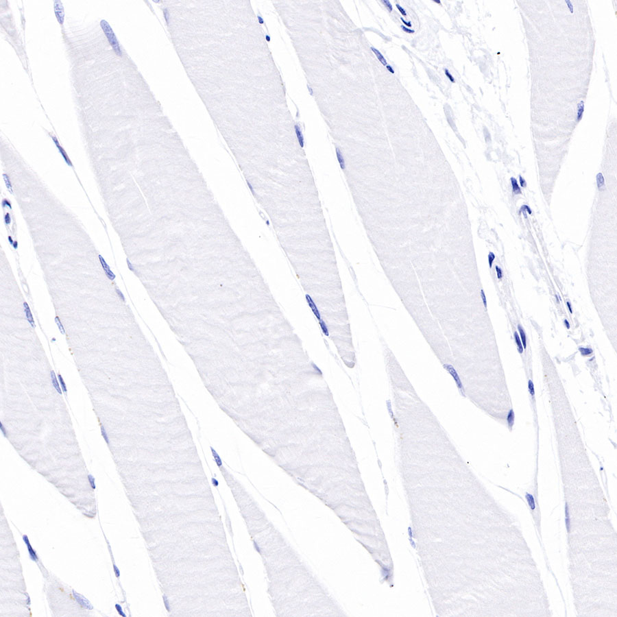
- Negative control: IHC shows negative staining in paraffin-embedded human skeletal muscle. Anti-CD138 antibody was used at 1/1000 dilution, followed by a HRP Polymer for Mouse & Rabbit IgG (ready to use). Counterstained with hematoxylin. Heat mediated antigen retrieval with Tris/EDTA buffer pH9.0 was performed before commencing with IHC staining protocol.
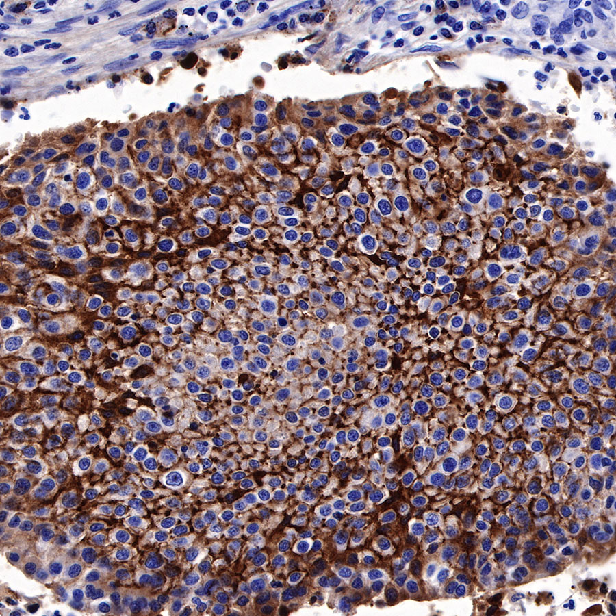
- IHC shows positive staining in paraffin-embedded human lung squamous cell carcinoma. Anti-CD138 antibody was used at 1/1000 dilution, followed by a HRP Polymer for Mouse & Rabbit IgG (ready to use). Counterstained with hematoxylin. Heat mediated antigen retrieval with Tris/EDTA buffer pH9.0 was performed before commencing with IHC staining protocol.
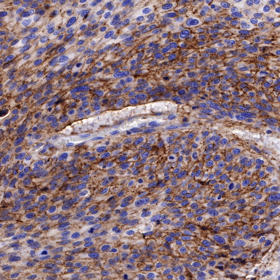
- IHC shows positive staining in paraffin-embedded human cervical squamous cell carcinoma. Anti-CD138 antibody was used at 1/1000 dilution, followed by a HRP Polymer for Mouse & Rabbit IgG (ready to use). Counterstained with hematoxylin. Heat mediated antigen retrieval with Tris/EDTA buffer pH9.0 was performed before commencing with IHC staining protocol.
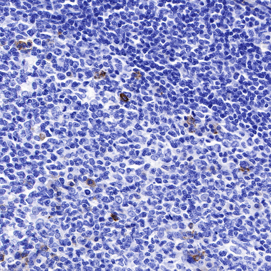
- IHC shows positive staining in paraffin-embedded human tonsil. Anti-CD138 antibody was used at 1/1000 dilution, followed by a HRP Polymer for Mouse & Rabbit IgG (ready to use). Counterstained with hematoxylin. Heat mediated antigen retrieval with Tris/EDTA buffer pH9.0 was performed before commencing with IHC staining protocol.
- IHC shows positive staining in paraffin-embedded human tonsil. Anti-CD138 antibody was used at 1/1000 dilution, followed by a HRP Polymer for Mouse & Rabbit IgG (ready to use). Counterstained with hematoxylin. Heat mediated antigen retrieval with Tris/EDTA buffer pH9.0 was performed before commencing with IHC staining protocol.
- IHC shows positive staining in paraffin-embedded human placenta. Anti-CD138 antibody was used at 1/1000 dilution, followed by a HRP Polymer for Mouse & Rabbit IgG (ready to use). Counterstained with hematoxylin. Heat mediated antigen retrieval with Tris/EDTA buffer pH9.0 was performed before commencing with IHC staining protocol.
- IHC shows positive staining in paraffin-embedded human colon. Anti-CD138 antibody was used at 1/1000 dilution, followed by a HRP Polymer for Mouse & Rabbit IgG (ready to use). Counterstained with hematoxylin. Heat mediated antigen retrieval with Tris/EDTA buffer pH9.0 was performed before commencing with IHC staining protocol.
免疫细胞化学
ICC shows positive staining in A431 cells. Anti-CD138 antibody was used at 1/25 dilution (Green) and incubated overnight at 4°C. Goat polyclonal Antibody to Rabbit IgG - H&L (Alexa Fluor® 488) was used as secondary antibody at 1/1000 dilution. The cells were fixed with 100% ice-cold methanol and permeabilized with 0.1% PBS-Triton X-100. Nuclei were counterstained with DAPI (Blue).
Negative control:ICC shows negative staining in Jurkat cells. Anti-CD138 antibody was used at 1/25 dilution and incubated overnight at 4°C. Goat polyclonal Antibody to Rabbit IgG - H&L (Alexa Fluor® 488) was used as secondary antibody at 1/1000 dilution. The cells were fixed with 100% ice-cold methanol and permeabilized with 0.1% PBS-Triton X-100. Nuclei were counterstained with DAPI (Blue).

评论(0)