


 大包装询价
大包装询价 产品介绍 评论(0)
宿主来源
Rabbit抗原名称
BOB-1分子别名
POU2AF1, OBF-1, OCA-B, OCT-binding factor 1免疫原
Synthetic Peptide细胞定位
NucleusAccession
Q16633克隆号
SDT-263-55抗体类型
Rabbit mAb应用
IHC-P, ICC, WB反应种属 ?
Hu, Ms, Rt纯化方式
Protein A浓度
0.125 mg/ml性状
Liquid缓冲体系
PBS, 40% Glycerol, 0.05% BSA, 0.03% Proclin 300
储存条件
12 months from date of receipt / reconstitution, -20 °C as supplied
| 应用 | 稀释度 |
|---|---|
| IHC-P | 1:500 |
| WB | 1:5000 |
| ICC | 1:125 |
BOB-1 interacts with the sequence-specific DNA-binding POU transcription factors (named after the founding family members PIT1, OCT1/2, and UNC86), the ubiquitously expressed OCT1 (POU2F1) and lymphoid-specific OCT2 (POU2F2). As a transcriptional co-activator, BOB-1 itself does not bind DNA but is rather recruited into transcriptional regulation via interaction with DNA-bound POU-domain transcription factors OCT1 and OCT2. The POU-domain is a unique bipartite structure allowing DNA recognition with remarkable flexibility .
免疫印迹
 WB result of BOB-1 Rabbit mAb Primary antibody: BOB-1 Rabbit mAb at 1/5000 dilution Lane 1: HeLa whole cell lysate 20 µg Lane 2: Raji whole cell lysate 20 µg Lane 3: Ramos whole cell lysate 20 µg Lane 4: Daudi whole cell lysate 20 µg Negative control: HeLa whole cell lysate Secondary antibody: Goat Anti-Rabbit IgG, (H+L), HRP conjugated at 1/10000 dilution Predicted MW: 27 kDa Observed MW: 37 kDa
免疫组化
 IHC shows positive staining in paraffin-embedded human tonsil. Anti-BOB-1 antibody was used at 1/500 dilution, followed by a HRP Polymer for Mouse & Rabbit IgG (ready to use). Counterstained with hematoxylin. Heat mediated antigen retrieval with Tris/EDTA buffer pH9.0 was performed before commencing with IHC staining protocol.
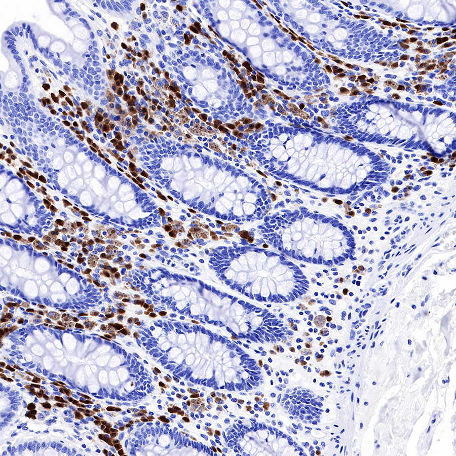 IHC shows positive staining in paraffin-embedded human colon. Anti-BOB-1 antibody was used at 1/500 dilution, followed by a HRP Polymer for Mouse & Rabbit IgG (ready to use). Counterstained with hematoxylin. Heat mediated antigen retrieval with Tris/EDTA buffer pH9.0 was performed before commencing with IHC staining protocol.
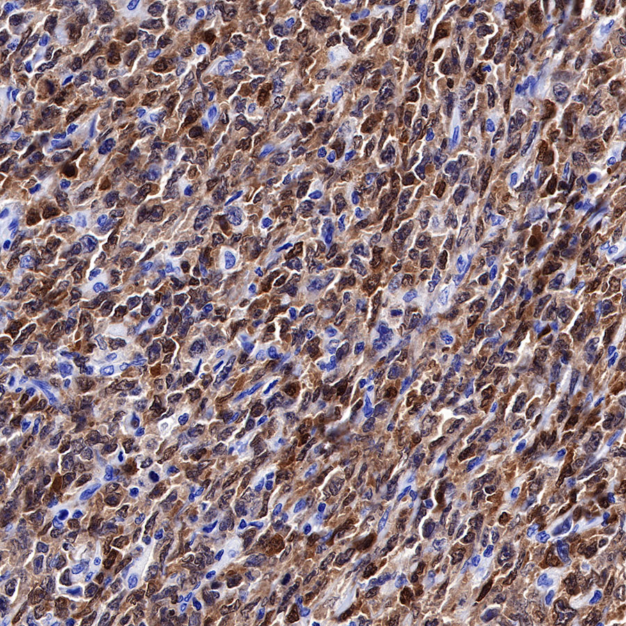 IHC shows positive staining in paraffin-embedded human diffuse large B-cell lymphoma. Anti-BOB-1 antibody was used at 1/500 dilution, followed by a HRP Polymer for Mouse & Rabbit IgG (ready to use). Counterstained with hematoxylin. Heat mediated antigen retrieval with Tris/EDTA buffer pH9.0 was performed before commencing with IHC staining protocol.
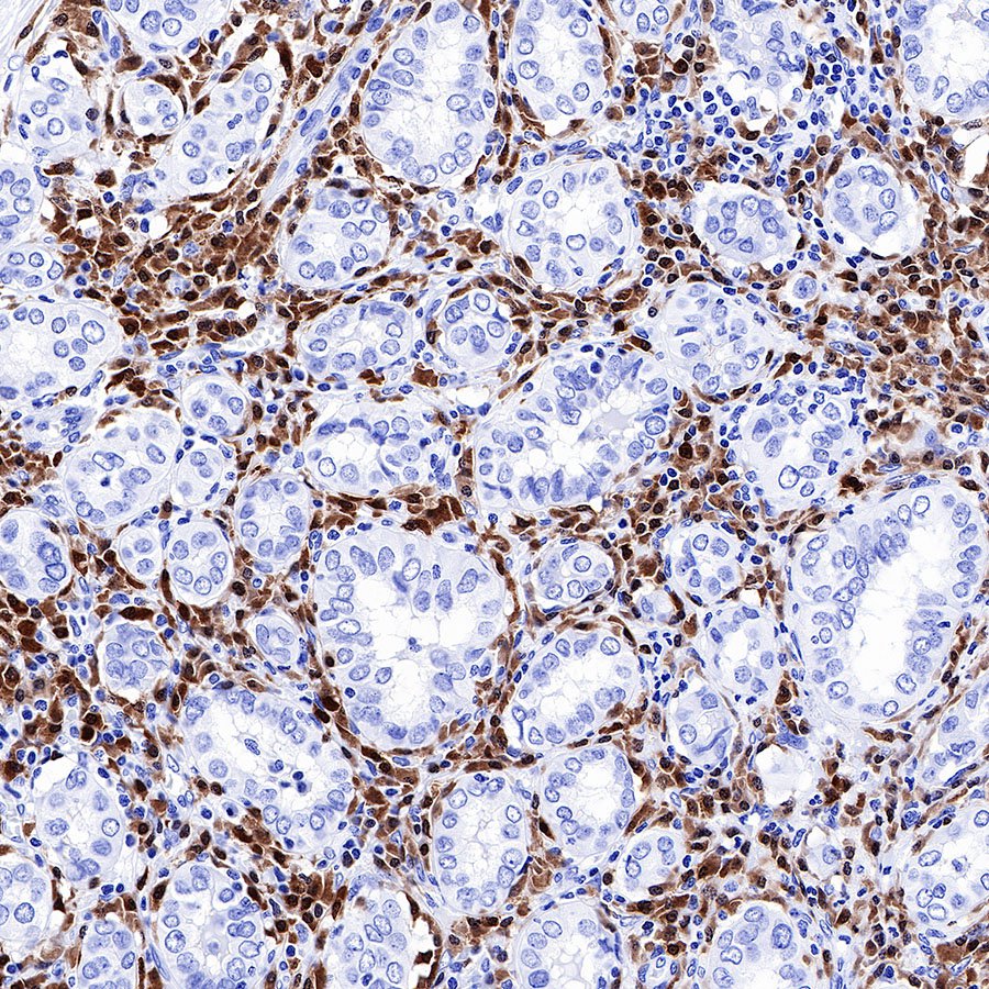 IHC shows positive staining in paraffin-embedded human thyroid cancer. Anti-BOB-1 antibody was used at 1/500 dilution, followed by a HRP Polymer for Mouse & Rabbit IgG (ready to use). Counterstained with hematoxylin. Heat mediated antigen retrieval with Tris/EDTA buffer pH9.0 was performed before commencing with IHC staining protocol.
 IHC shows positive staining in paraffin-embedded human diffuse large B-cell lymphoma. Anti-BOB-1 antibody was used at 1/500 dilution, followed by a HRP Polymer for Mouse & Rabbit IgG (ready to use). Counterstained with hematoxylin. Heat mediated antigen retrieval with Tris/EDTA buffer pH9.0 was performed before commencing with IHC staining protocol.
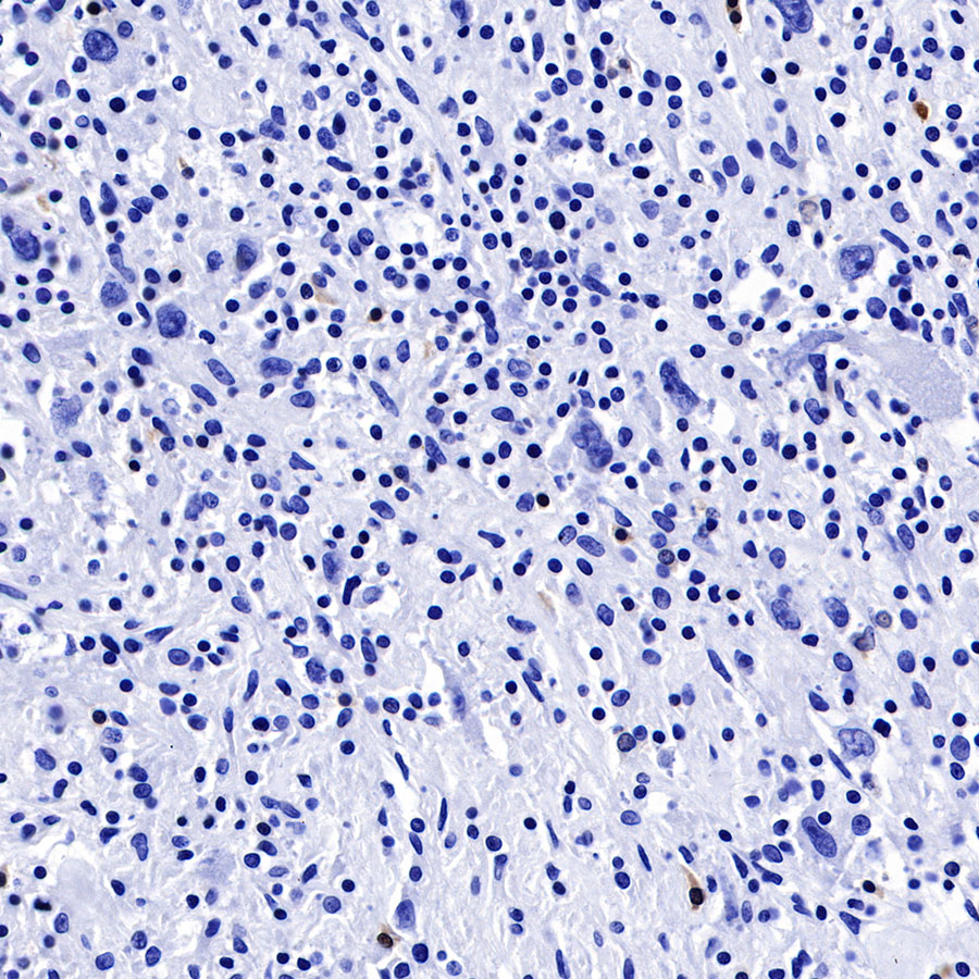 Negative control: IHC shows negative staining in paraffin-embedded human Hodgkin's lymphoma. Anti-BOB-1 antibody was used at 1/500 dilution, followed by a HRP Polymer for Mouse & Rabbit IgG (ready to use). Counterstained with hematoxylin. Heat mediated antigen retrieval with Tris/EDTA buffer pH9.0 was performed before commencing with IHC staining protocol.
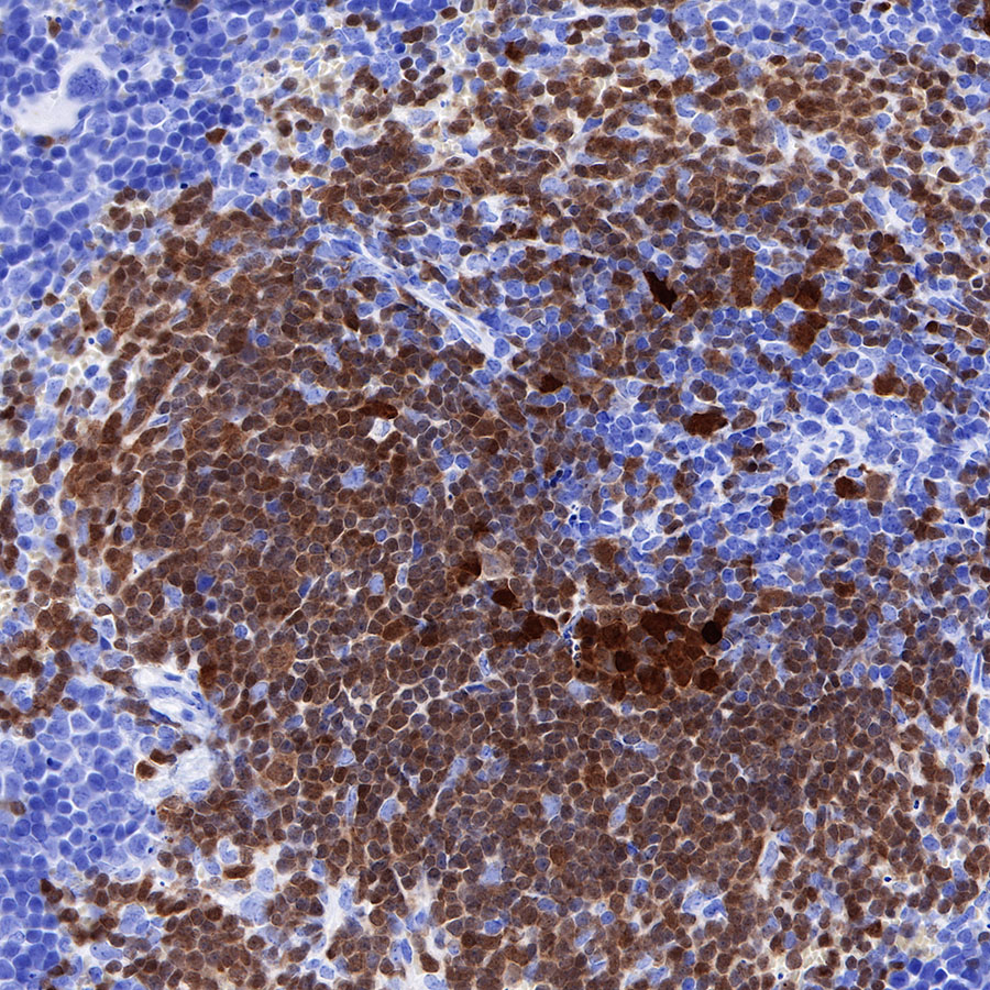 IHC shows positive staining in paraffin-embedded mouse spleen. Anti-BOB-1 antibody was used at 1/500 dilution, followed by a HRP Polymer for Mouse & Rabbit IgG (ready to use). Counterstained with hematoxylin. Heat mediated antigen retrieval with Tris/EDTA buffer pH9.0 was performed before commencing with IHC staining protocol.
 IHC shows positive staining in paraffin-embedded rat spleen. Anti-BOB-1 antibody was used at 1/500 dilution, followed by a HRP Polymer for Mouse & Rabbit IgG (ready to use). Counterstained with hematoxylin. Heat mediated antigen retrieval with Tris/EDTA buffer pH9.0 was performed before commencing with IHC staining protocol.
免疫细胞化学

ICC shows positive staining in Ramos cells. Anti-BOB-1 antibody was used at 1/125 dilution (Green) and incubated overnight at 4°C. Goat polyclonal Antibody to Rabbit IgG - H&L (Alexa Fluor® 488) was used as secondary antibody at 1/1000 dilution. The cells were fixed with 4% PFA and permeabilized with 0.1% PBS-Triton X-100. Nuclei were counterstained with DAPI (Blue).
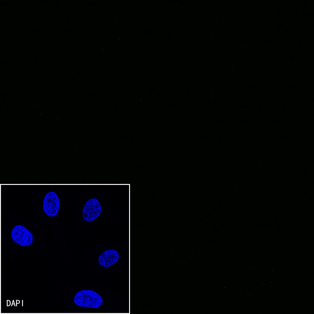
Negative control:ICC shows negative staining in HeLa cells. Anti-BOB-1 antibody was used at 1/125 dilution and incubated overnight at 4°C. Goat polyclonal Antibody to Rabbit IgG - H&L (Alexa Fluor® 488) was used as secondary antibody at 1/1000 dilution. The cells were fixed with 4% PFA and permeabilized with 0.1% PBS-Triton X-100. Nuclei were counterstained with DAPI (Blue).

评论(0)