


PD-L1 Recombinant Rabbit mAb (SDT-R017)
PD-L1,CD274,B7-H,B7-H1,B7H1,PDCD1LG1,PDL1,PDCD1L1,CD27L receptor,T-cell activation antigen CD27,T14,Tumor necrosis factor receptor superfamily member 7,PDCD1 ligand 1,Programmed death ligand 1,hPD-L1,Programmed cell death 1 ligand 1
货号: S0B2134
- 价格: ¥600
- 规格:
- 数量:
 大包装询价
大包装询价 产品介绍 评论(0)
宿主来源
Rabbit抗原名称
PD-L1分子别名
Programmed cell death 1 ligand 1, CD274, B7-H1免疫原
N/A细胞定位
MembraneAccession
Q9NZQ7克隆号
SDT-R017抗体类型
Rabbit mAb应用
IHC-P, WB, IF反应种属 ?
Hu纯化方式
Protein A浓度
0.5 mg/ml性状
Liquid缓冲体系
PBS, 40% Glycerol, 0.05% BSA, 0.03% Proclin 300储存条件
12 months from date of receipt / reconstitution, -20 °C as supplied
| 应用 | 稀释度 |
|---|---|
| IHC-P | 1:500 |
| WB | 1:1000 |
| IF | 1:100 |
Programmed death-ligand 1 (PD-L1) also known as cluster of differentiation 274 (CD274) or B7 homolog 1 (B7-H1) is a protein that in humans is encoded by the CD274 gene. Programmed death-ligand 1 (PD-L1) is a 40kDa type 1 transmembrane protein that has been speculated to play a major role in suppressing the adaptive arm of immune systems during particular events such as pregnancy, tissue allografts, autoimmune disease and other disease states such as hepatitis. The binding of PD-L1 to the inhibitory checkpoint molecule PD-1 transmits an inhibitory signal based on interaction with phosphatases (SHP-1 or SHP-2) via Immunoreceptor Tyrosine-Based Switch Motif (ITSM). This reduces the proliferation of antigen-specific T-cells in lymph nodes, while simultaneously reducing apoptosis in regulatory T cells (anti-inflammatory, suppressive T cells) - further mediated by a lower regulation of the gene Bcl-2. Upregulation of PD-L1 on immune cells (especially myeloid cells) can also lead to formation of an immunosuppressive environment in a highly localized manner that also allow the cancer cells to proliferate.
免疫印迹

WB result of PD-L1 Rabbit mAb
Primary antibody: PD-L1 Rabbit mAb at 1/1000 dilution
Lane 1: Untreated A549 whole cell lysate 20 µg
Lane 2: A549 treated with IFNγ (100 ng/ml, 48 hr) whole cell lysate 20 µg
Negative control: Untreated A549 whole cell lysate
Secondary antibody: Goat Anti-Rabbit IgG, (H+L), HRP conjugated at 1/10000 dilution
Predicted MW: 33 kDa
Observed MW: 40~50 kDa
Exposure time: 180s
免疫组化
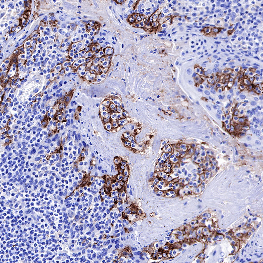
IHC shows positive staining in paraffin-embedded human tonsil. Anti-PD-L1 antibody was used at 1/500 dilution, followed by a HRP Polymer for Mouse & Rabbit IgG (ready to use). Counterstained with hematoxylin. Heat mediated antigen retrieval with Tris/EDTA buffer pH9.0 was performed before commencing with IHC staining protocol.
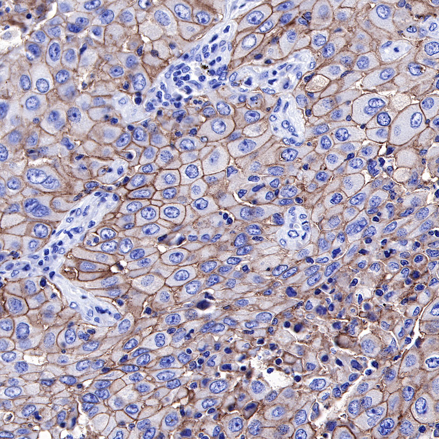
IHC shows positive staining in paraffin-embedded human lung cancer. Anti-PD-L1 antibody was used at 1/500 dilution, followed by a HRP Polymer for Mouse & Rabbit IgG (ready to use). Counterstained with hematoxylin. Heat mediated antigen retrieval with Tris/EDTA buffer pH9.0 was performed before commencing with IHC staining protocol.
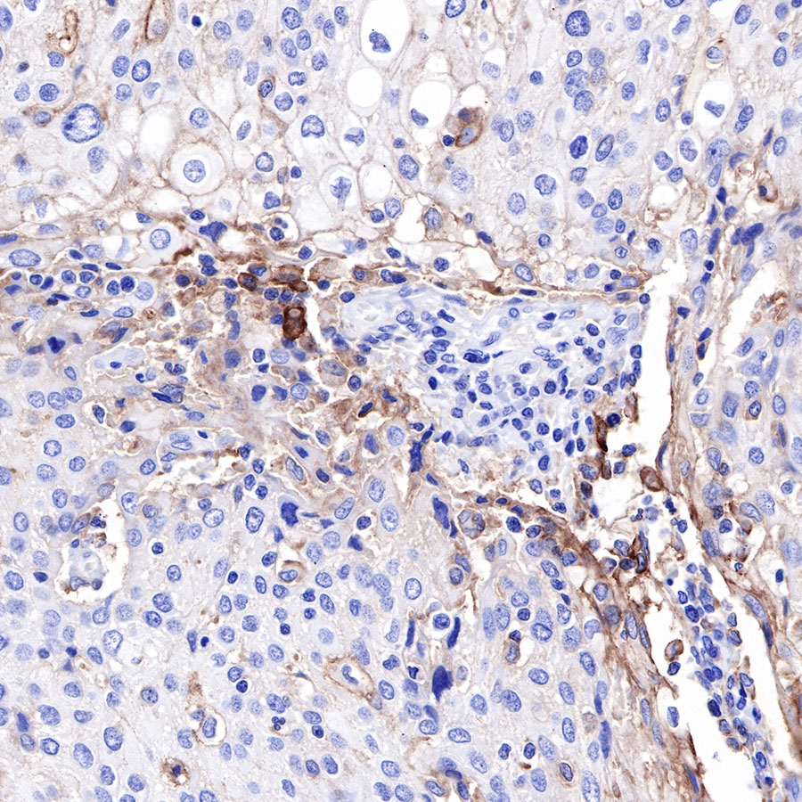
IHC shows positive staining in paraffin-embedded human cervical carcinoma. Anti-PD-L1 antibody was used at 1/500 dilution, followed by a HRP Polymer for Mouse & Rabbit IgG (ready to use). Counterstained with hematoxylin. Heat mediated antigen retrieval with Tris/EDTA buffer pH9.0 was performed before commencing with IHC staining protocol.
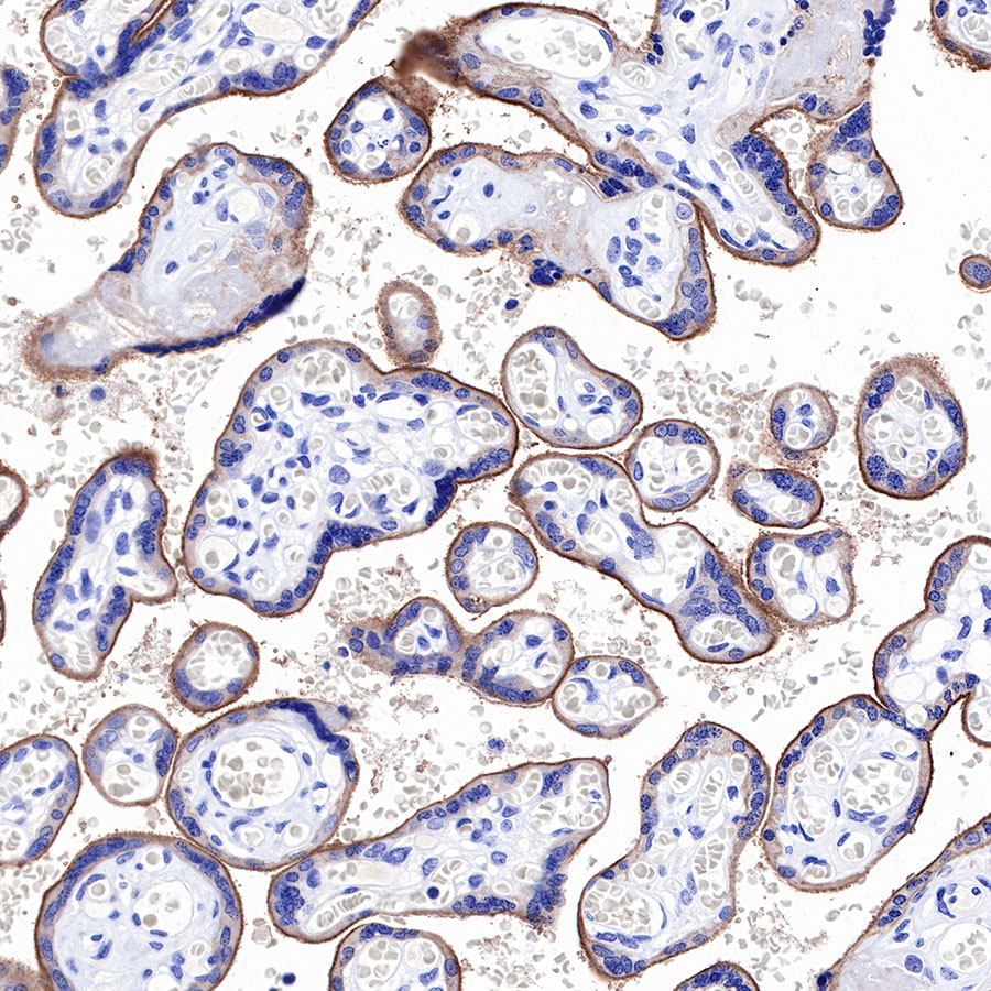
IHC shows positive staining in paraffin-embedded human placenta. Anti-PD-L1 antibody was used at 1/500 dilution, followed by a HRP Polymer for Mouse & Rabbit IgG (ready to use). Counterstained with hematoxylin. Heat mediated antigen retrieval with Tris/EDTA buffer pH9.0 was performed before commencing with IHC staining protocol.
免疫荧光
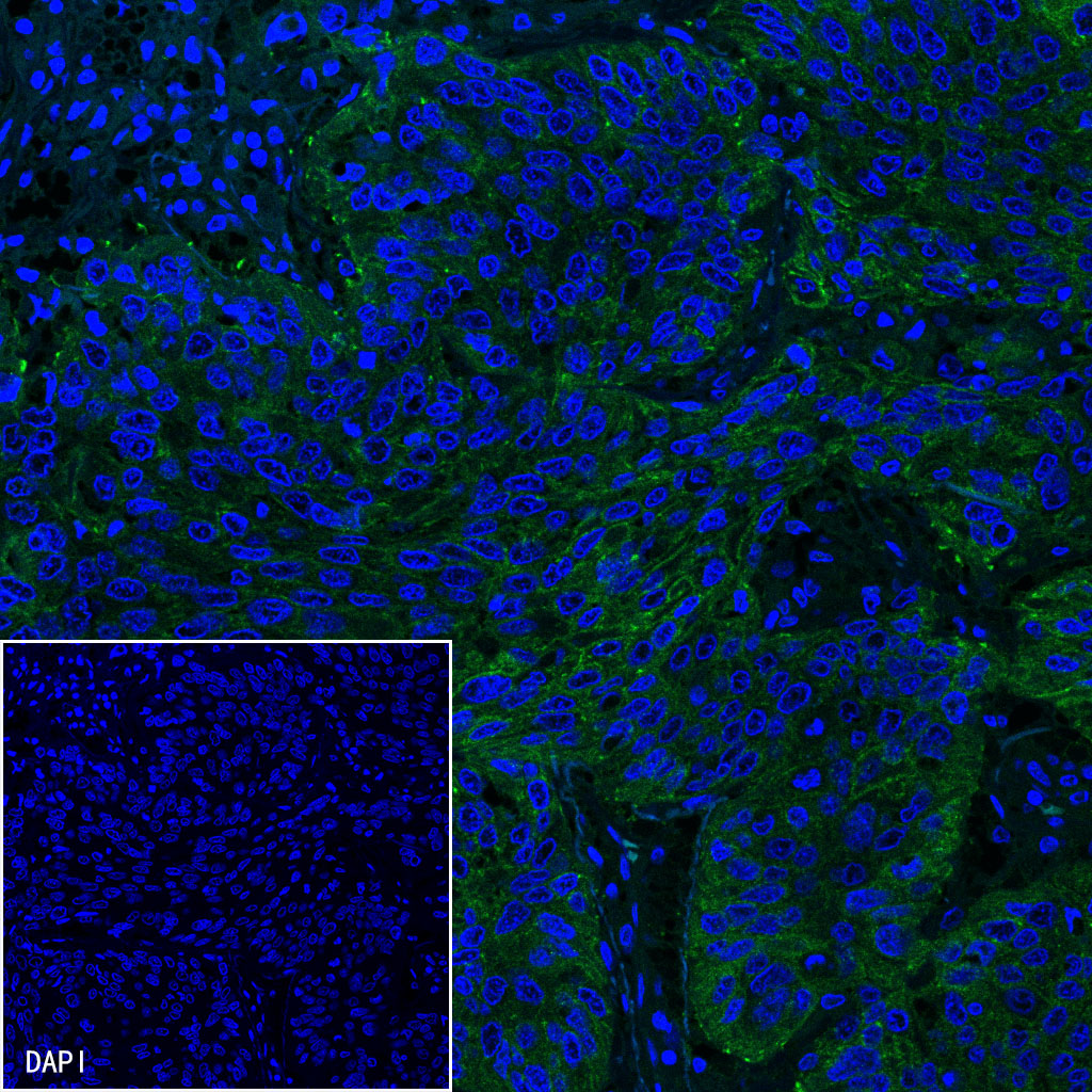
IF shows positive staining in paraffin-embedded human lung squamous cell carcainoma. Anti-PD-L1 antibody was used at 1/100 dilution (Green) and incubated overnight at 4°C. Goat polyclonal Antibody to Rabbit IgG - H&L (Alexa Fluor® 488) was used as secondary antibody at 1/1000 dilution. Counterstained with DAPI (Blue). Heat mediated antigen retrieval with EDTA buffer pH9.0 was performed before commencing with IF staining protocol.

评论(0)