


 申请试用
申请试用 产品介绍 评论(0)
宿主来源
Rabbit抗原名称
Cytokeratin 19 fragment (CY211)分子别名
CYFRA 21-1免疫原
Recombinant Protein细胞定位
CytoplasmAccession
P08727克隆号
SDT-197-28抗体类型
Rabbit mAb应用
IHC-P ? ,ICC ? ,WB反应种属 ?
Hu纯化方式
Protein A浓度
1 mg/ml性状
Liquid缓冲体系
PBS
储存条件
12 months from date of receipt, 4°C as supplied
| 应用 | 稀释度 |
|---|---|
| WB | 1:1000-1:5000 |
| IHC-P | 1:1000 |
| ICC | 1:250 |
Cytokeratin 19 fragment (CYFRA 21-1) is a fragment of cytokeratin 19 that is typically associated with epithelial cell cancers, including NSCLC, and is typically associated with the SQLC type. Since cytokeratins are structural proteins of keratin-containing intermediate filaments found in the epithelial cells, their degradation produces soluble fragments which are measurable in the blood of lung cancer patients as a tumor marker. Other examples of cytokeratin fragments include tissue polypeptide antigen and tissue polypeptide-specific antigen. CYFRA 21-1 is known to be correlated with disease response and the prognosis of lung cancer but cannot be used to differentiate cancer patients from patients with respiratory diseases.
免疫印迹
WB result of Cytokeratin 19 fragment (CY211) Rabbit mAb
Primary antibody: Cytokeratin 19 fragment (CY211) Rabbit mAb at 1/5000 dilution
Lane 1: HeLa whole cell lysate 20 µg
Lane 2: HT-29 whole cell lysate 20 µg
Lane 3: HCT 116 whole cell lysate 20 µg
Lane 4: MCF7 whole cell lysate 20 µg
Lane 5: HepG2 whole cell lysate 20 µg
Lane 6: SK-OV-3 whole cell lysate 20 µg
Negative control: HeLa whole cell lysate
Secondary antibody: Goat Anti-Rabbit IgG, (H+L), HRP conjugated at 1/10000 dilution
Predicted MW: 40 kDa
Observed MW: 38, 40 kDa
免疫组化
IHC shows positive staining in paraffin-embedded human colon cancer. Anti-Cytokeratin 19 fragment (CY211) antibody was used at 1/1000 dilution, followed by a HRP Polymer for Mouse & Rabbit IgG (ready to use). Counterstained with hematoxylin. Heat mediated antigen retrieval with Tris/EDTA buffer pH9.0 was performed before commencing with IHC staining protocol.
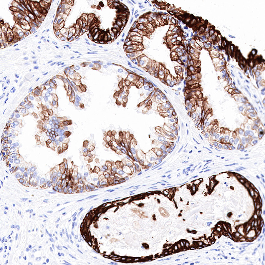
IHC shows positive staining in paraffin-embedded human colon. Anti-Cytokeratin 19 fragment (CY211) antibody was used at 1/1000 dilution, followed by a HRP Polymer for Mouse & Rabbit IgG (ready to use). Counterstained with hematoxylin. Heat mediated antigen retrieval with Tris/EDTA buffer pH9.0 was performed before commencing with IHC staining protocol.
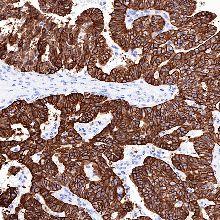
IHC shows positive staining in paraffin-embedded human lung adenocarcinoma. Anti-Cytokeratin 19 fragment (CY211) antibody was used at 1/1000 dilution, followed by a HRP Polymer for Mouse & Rabbit IgG (ready to use). Counterstained with hematoxylin. Heat mediated antigen retrieval with Tris/EDTA buffer pH9.0 was performed before commencing with IHC staining protocol.
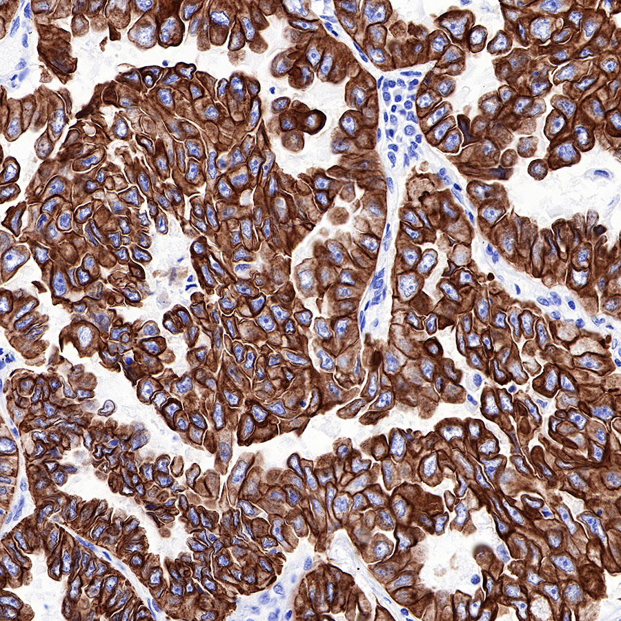
IHC shows positive staining in paraffin-embedded human ovarian carcinoma. Anti-Cytokeratin 19 fragment (CY211) antibody was used at 1/1000 dilution, followed by a HRP Polymer for Mouse & Rabbit IgG (ready to use). Counterstained with hematoxylin. Heat mediated antigen retrieval with Tris/EDTA buffer pH9.0 was performed before commencing with IHC staining protocol.
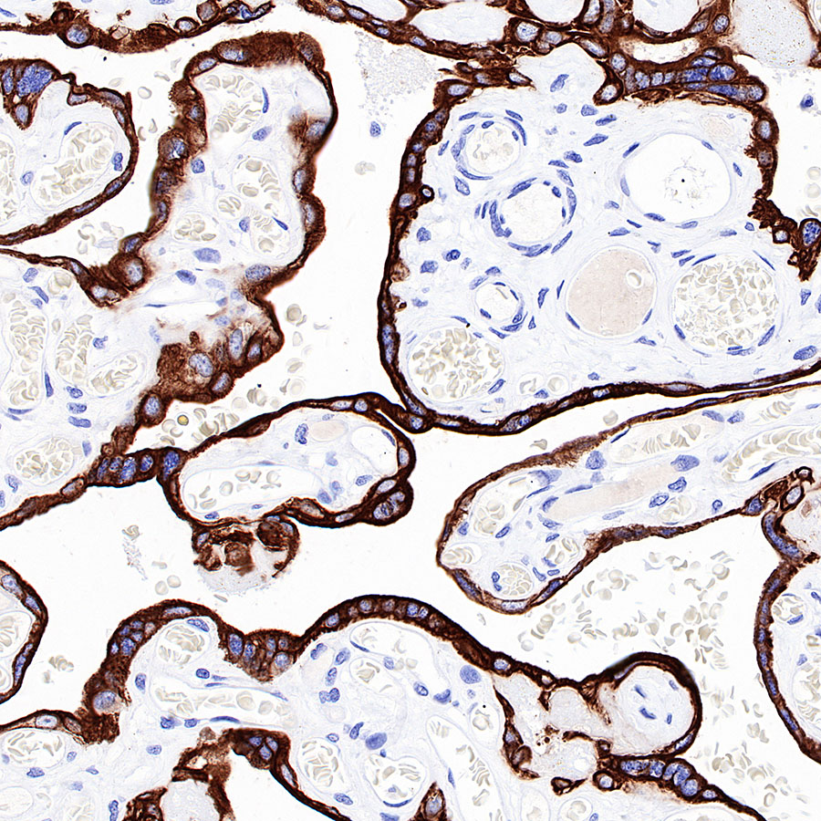
IHC shows positive staining in paraffin-embedded human placenta. Anti-Cytokeratin 19 fragment (CY211) antibody was used at 1/1000 dilution, followed by a HRP Polymer for Mouse & Rabbit IgG (ready to use). Counterstained with hematoxylin. Heat mediated antigen retrieval with Tris/EDTA buffer pH9.0 was performed before commencing with IHC staining protocol.
IHC shows positive staining in paraffin-embedded human prostatic hyperplasia. Anti-Cytokeratin 19 fragment (CY211) antibody was used at 1/1000 dilution, followed by a HRP Polymer for Mouse & Rabbit IgG (ready to use). Counterstained with hematoxylin. Heat mediated antigen retrieval with Tris/EDTA buffer pH9.0 was performed before commencing with IHC staining protocol.
免疫细胞化学
ICC shows positive staining in MCF7 cells. Anti-Cytokeratin 19 fragment (CY211) antibody was used at 1/500 dilution (Green) and incubated overnight at 4°C. Goat polyclonal Antibody to Rabbit IgG - H&L (Alexa Fluor® 488) was used as secondary antibody at 1/1000 dilution. The cells were fixed with 100% ice-cold methanol and permeabilized with 0.1% PBS-Triton X-100. Nuclei were counterstained with DAPI (Blue).Counterstain with tubulin (Red).

评论(0)