


 大包装询价
大包装询价 产品介绍 评论(0)
宿主来源
Rabbit抗原名称
S100B分子别名
Protein S100-B, S-100 protein beta chain免疫原
Recombinant Protein细胞定位
Nucleus, CytoplasmAccession
P04271克隆号
SDT-093-108抗体类型
Rabbit mAb应用
IHC-P, WB, IF反应种属 ?
Hu, Ms, Rt纯化方式
Protein A浓度
1 mg/ml性状
Liquid缓冲体系
PBS
储存条件
12 months from date of receipt, 4°C as supplied
| 应用 | 稀释度 |
|---|---|
| WB | 1:500 |
| IHC-P | 1:2500 |
| IF | 1:1250 |
S100B is a Ca2+-binding protein, which is secreted from astrocytes. S100B is widely expressed in astrocytes, certain neuronal populations, Schwann cells, myeloid-derived cells, and a few other cell types. S100B is released in the extracellular space in response to glutamate, serotonin, TNF, IL-1β, beta-amyloid peptides, and lysophosphatidic acid. Extracellular S100B exerts both paracrine and autocrine effects on neurons and glia100, through RAGE, and possibly other receptors. The effect of S100B is concentration dependent and can be either trophic or toxic. In humans, high levels of extracellular S100B have been detected under various clinical conditions, for example, brain trauma, ischemia and neurodegenerative, and inflammatory and psychiatric diseases.
免疫印迹
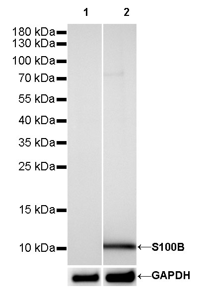
WB result of S100B Rabbit mAb
Primary antibody: S100B Rabbit mAb at 1/500 dilution
Lane 1: T-47D whole cell lysate 5 µg
Lane 2: SK-MEL-28 whole cell lysate 5 µg
Negative control: T-47D whole cell lysateSecondary antibody: Goat Anti-Rabbit IgG, (H+L), HRP conjugated at 1/10000 dilution
Predicted MW: 10 kDa
Observed MW: 10 kDa
Exposure time: 30s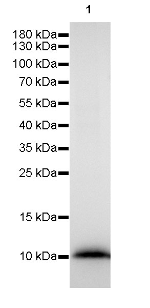
WB result of S100B Rabbit mAb
Primary antibody: S100B Rabbit mAb at 1/500 dilution
Lane 1: rat brain whole cell lysate 5 µg
Secondary antibody: Goat Anti-Rabbit IgG, (H+L), HRP conjugated at 1/10000 dilution
Predicted MW: 10 kDa
Observed MW: 10 kDa
Exposure time: 16s
免疫组化
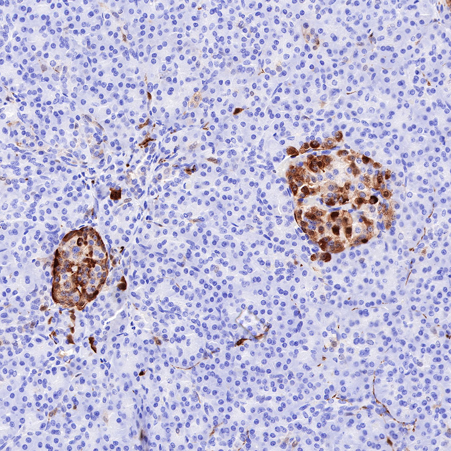
IHC shows positive staining in paraffin-embedded human pancreas. Anti-S100B antibody was used at 1/2500 dilution, followed by a HRP Polymer for Mouse & Rabbit IgG (ready to use). Counterstained with hematoxylin. Heat mediated antigen retrieval with Tris/EDTA buffer pH9.0 was performed before commencing with IHC staining protocol.
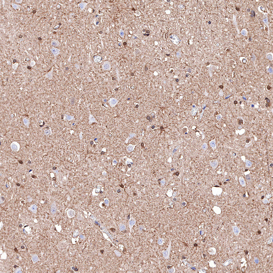
IHC shows positive staining in paraffin-embedded human cerebral cortex. Anti-S100B antibody was used at 1/2500 dilution, followed by a HRP Polymer for Mouse & Rabbit IgG (ready to use). Counterstained with hematoxylin. Heat mediated antigen retrieval with Tris/EDTA buffer pH9.0 was performed before commencing with IHC staining protocol.
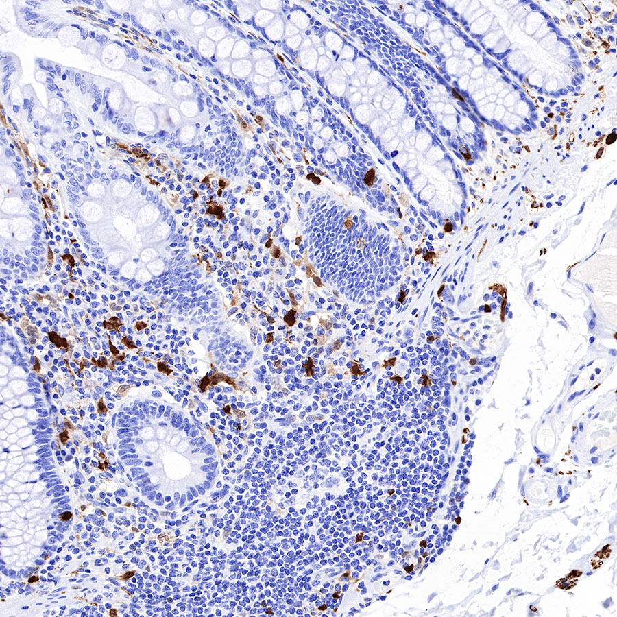
IHC shows positive staining in paraffin-embedded human colon. Anti-S100B antibody was used at 1/2500 dilution, followed by a HRP Polymer for Mouse & Rabbit IgG (ready to use). Counterstained with hematoxylin. Heat mediated antigen retrieval with Tris/EDTA buffer pH9.0 was performed before commencing with IHC staining protocol.
IHC shows positive staining in paraffin-embedded mouse cerebral cortex. Anti-S100B antibody was used at 1/2500 dilution, followed by a HRP Polymer for Mouse & Rabbit IgG (ready to use). Counterstained with hematoxylin. Heat mediated antigen retrieval with Tris/EDTA buffer pH9.0 was performed before commencing with IHC staining protocol.

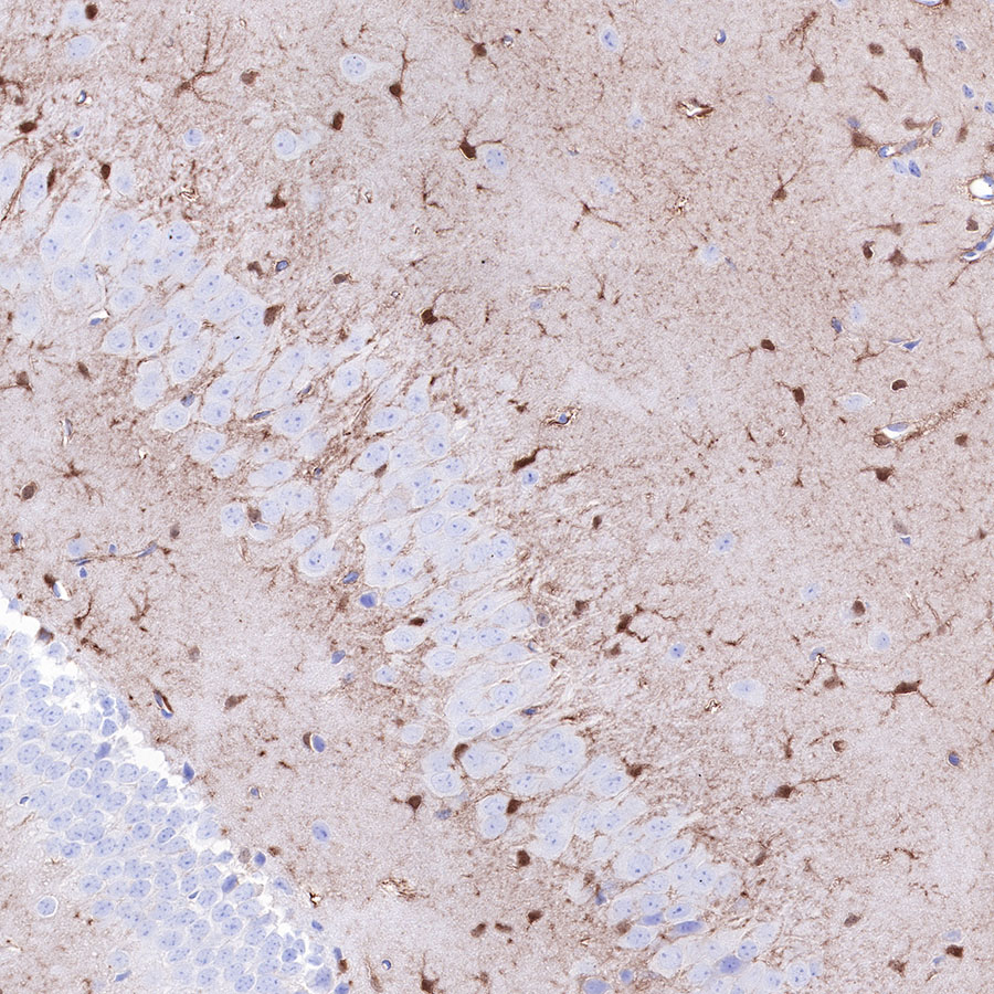
IHC shows positive staining in paraffin-embedded mouse cerebellum. Anti-S100B antibody was used at 1/2500 dilution, followed by a HRP Polymer for Mouse & Rabbit IgG (ready to use). Counterstained with hematoxylin. Heat mediated antigen retrieval with Tris/EDTA buffer pH9.0 was performed before commencing with IHC staining protocol.
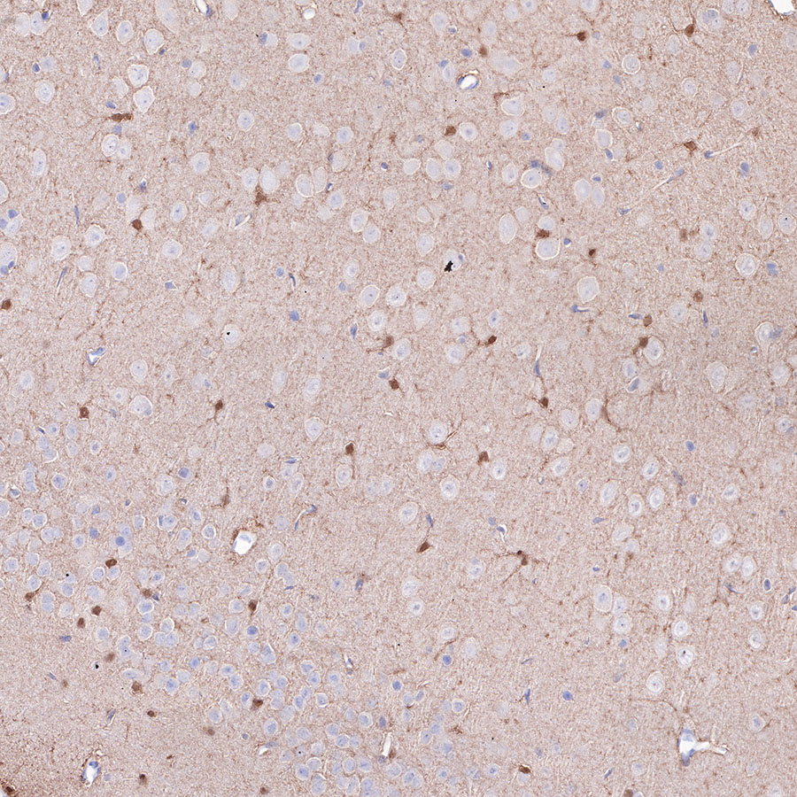
IHC shows positive staining in paraffin-embedded rat cerebral cortex. Anti-S100B antibody was used at 1/2500 dilution, followed by a HRP Polymer for Mouse & Rabbit IgG (ready to use). Counterstained with hematoxylin. Heat mediated antigen retrieval with Tris/EDTA buffer pH9.0 was performed before commencing with IHC staining protocol.
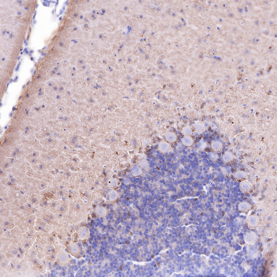
IHC shows positive staining in paraffin-embedded rat cerebellum. Anti-S100B antibody was used at 1/2500 dilution, followed by a HRP Polymer for Mouse & Rabbit IgG (ready to use). Counterstained with hematoxylin. Heat mediated antigen retrieval with Tris/EDTA buffer pH9.0 was performed before commencing with IHC staining protocol.
免疫荧光
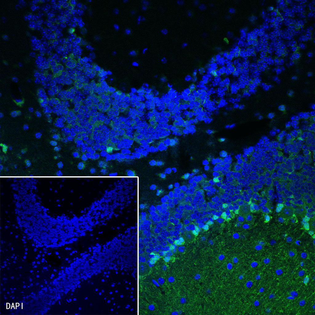
IF shows positive staining in paraffin-embedded mouse cerebellum. Anti-S100B antibody was used at 1/1250 dilution (Green) and incubated overnight at 4°C. Goat polyclonal Antibody to Rabbit IgG - H&L (Alexa Fluor® 488) was used as secondary antibody at 1/1000 dilution. Counterstained with DAPI (Blue). Heat mediated antigen retrieval with Tris/EDTA buffer pH9.0 was performed before commencing with IF staining protocol.

评论(0)