




 分享
分享产品介绍 引用文献(1) 评论(0)
宿主来源
Rabbit抗原名称
E-Cadherin分子别名
Cadherin-1, CD324细胞定位
Cell membraneAccession
P12830克隆号
SDT-R088抗体类型
Rabbit mAb反应种属 ?
Hu纯化方式
Protein A浓度
0.25 mg/ml性状
Liquid缓冲体系
PBS, 40% Glycerol, 0.05%BSA, 0.03% Proclin 300
储存条件
12 months from date of receipt / reconstitution, -20 °C as supplied
应用
IHC-P ?
ICC ?
WB
稀释度
应用 稀释度 IHC-P 1:1000 WB 1:1000-1:5000 ICC 1:100
E-cadherin is a transmembrane glycoprotein which connects epithelial cells together at adherens junctions. In normal cells, E-cadherin exerts its tumour suppressing role mainly by sequestering β-catenin from its binding to LEF (Lymphoid enhancer factor)/TCF (T cell factor) which serves the function of transcribing genes of the proliferative Wnt signaling pathway. Despite the ongoing debate on whether the loss of E-cadherin is the cause or effect of epithelial-mesenchymal transition (EMT), E-cadherin functional loss has frequently been associated with poor prognosis and survival in patients of various cancers. The dysregulation of E-cadherin expression that leads to carcinogenesis happens mostly at the epigenetic level but there are cases of genetic alterations as well. E-cadherin expression has been linked to the cellular functions of invasiveness reduction, growth inhibition, apoptosis, cell cycle arrest and differentiation.
免疫印迹
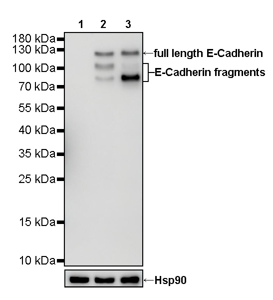
WB result of E-cadherin Rabbit mAb
Primary antibody: E-cadherin Rabbit mAb at 1/5000 dilution
Lane 1: HeLa whole cell lysate 20 µg
Lane 2: MCF7 whole cell lysate 20 µg
Lane 3: HT-29 whole cell lysate 20 µg
Negative control: HeLa whole cell lysate
Secondary antibody: Goat Anti-Rabbit IgG, (H+L), HRP conjugated at 1/10000 dilution
Predicted MW: 97 kDa
Observed MW: 80~125 kDa
免疫组化
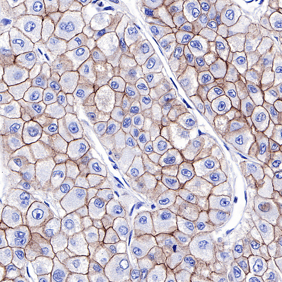
IHC shows positive staining in paraffin-embedded human hepatocellular carcinoma. Anti-E-cadherin antibody was used at 1/1000 dilution, followed by a HRP Polymer for Mouse & Rabbit IgG (ready to use). Counterstained with hematoxylin. Heat mediated antigen retrieval with Tris/EDTA buffer pH9.0 was performed before commencing with IHC staining protocol.
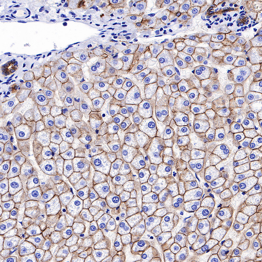
IHC shows positive staining in paraffin-embedded human liver. Anti-E-cadherin antibody was used at 1/1000 dilution, followed by a HRP Polymer for Mouse & Rabbit IgG (ready to use). Counterstained with hematoxylin. Heat mediated antigen retrieval with Tris/EDTA buffer pH9.0 was performed before commencing with IHC staining protocol.
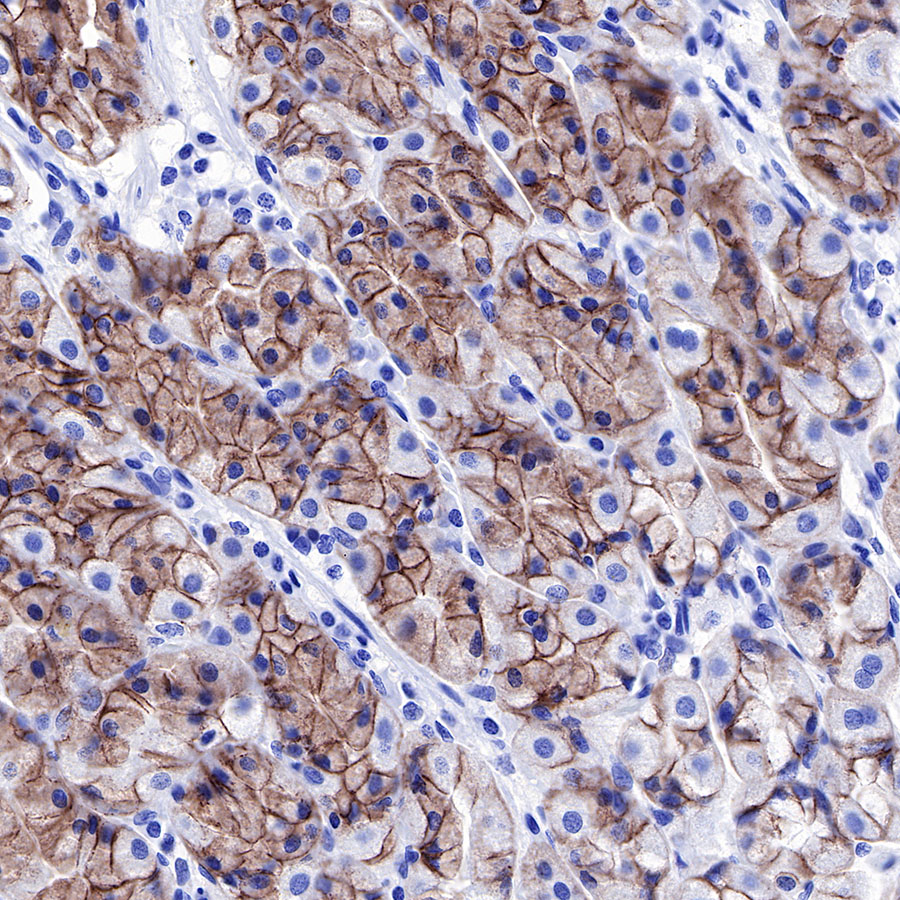
IHC shows positive staining in paraffin-embedded human stomach. Anti-E-cadherin antibody was used at 1/1000 dilution, followed by a HRP Polymer for Mouse & Rabbit IgG (ready to use). Counterstained with hematoxylin. Heat mediated antigen retrieval with Tris/EDTA buffer pH9.0 was performed before commencing with IHC staining protocol.
IHC shows positive staining in paraffin-embedded human pancreatic carcinoma. Anti-E-cadherin antibody was used at 1/1000 dilution, followed by a HRP Polymer for Mouse & Rabbit IgG (ready to use). Counterstained with hematoxylin. Heat mediated antigen retrieval with Tris/EDTA buffer pH9.0 was performed before commencing with IHC staining protocol.
免疫细胞化学
ICC shows positive staining in MCF7 cells (top panel) and negative staining in HeLa cells (below panel). Anti-E-Cadherin antibody was used at 1/100 dilution (Green) and incubated overnight at 4°C. Goat polyclonal Antibody to Rabbit IgG - H&L (Alexa Fluor® 488) was used as secondary antibody at 1/1000 dilution. The cells were fixed with 4% PFA and permeabilized with 0.1% PBS-Triton X-100. Nuclei were counterstained with DAPI (Blue). Counterstain with tubulin (Red).
引用文献(1)
- FN1+ macrophages fuel aggressive sarcomatoid differentiation and immunotherapy refractory outcome in clear cell renal cell carcinoma
Jiangting Cheng,Wenbin Jiang, Li Liu, Ziyang Xu, Yu Dong, Youqi Qiu, Yang Qu, Yu Xia, Siyuan Dai, Jianming Guo, Jiejie Xu
Oncogene. 2026 May 01 .
影响因子: 9.1
货号:S0B2112产品名称:S-RMab® E-Cadherin Recombinant Rabbit mAb (SDT-R088)

评论(0)