


 大包装询价
大包装询价 产品介绍 评论(0)
宿主来源
Rabbit抗原名称
Myoglobin分子别名
Mb,MB免疫原
Recombinant Protein细胞定位
CytoplasmAccession
P02144克隆号
SDT-097-10抗体类型
Rabbit mAb应用
ICFCM, IHC-P, WB, IF反应种属 ?
Hu, Ms, Rt纯化方式
Protein A浓度
1 mg/ml性状
Liquid缓冲体系
PBS储存条件
12 months from date of receipt, 4°C as supplied
| 应用 | 稀释度 |
|---|---|
| WB | 1:500 |
| IHC-P | 1:1000 |
| ICFCM | 1:250 |
| IF | 1:1000 |
Myoglobin is a protein that's found in your striated muscles, which includes skeletal muscles (the muscles attached to your bones and tendons) and heart muscles. Its main function is to supply oxygen to the cells in your muscles (myocytes). All cells in your body need oxygen in order to function. Myoglobin is distantly related to hemoglobin. Compared to hemoglobin, myoglobin has a higher affinity for oxygen and does not have cooperative binding with oxygen like hemoglobin does. In humans, myoglobin is only found in the bloodstream after muscle injury.
验证数据
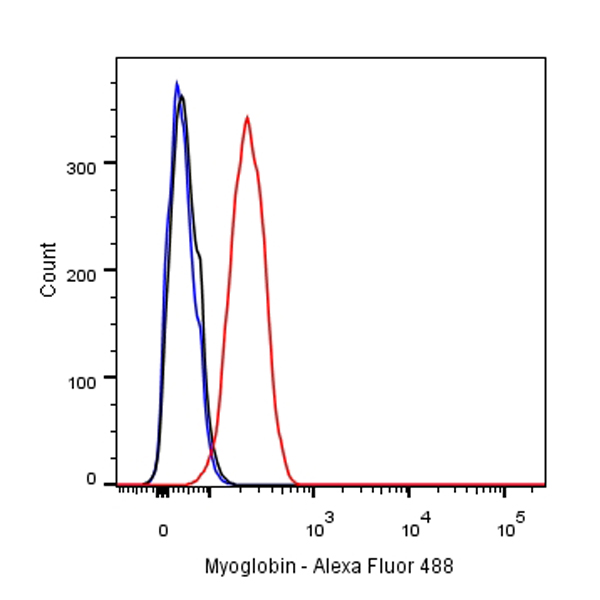
Flow cytometric analysis of SK-BR-3 cells labelling Myoglobin antibody at 1/250 dilution (0.1ug)/ (red) compared with a Rabbit monoclonal IgG (Black) isotype control and an unlabelled control (cells without incubation with primary antibody and secondary antibody) (Blue). Goat Anti-Rabbit IgG Alexa Fluor® 488 was used as the secondary antibody.
免疫印迹
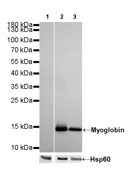
WB result of Myoglobin Rabbit mAb
Primary antibody: Myoglobin Rabbit mAb at 1/500 dilution
Lane 1: mouse brain lysate 20 µg
Lane 2: mouse heart lysate 20 µg
Lane 3: mouse skeletal muscle lysate 20 µg
Negative control: mouse brain lysateSecondary antibody: Goat Anti-Rabbit IgG, (H+L), HRP conjugated at 1/10000 dilution
Predicted MW: 17 kDa
Observed MW: 14 kDa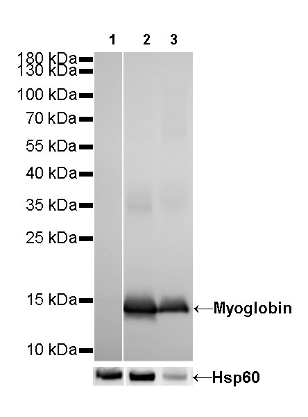
WB result of Myoglobin Rabbit mAb
Primary antibody: Myoglobin Rabbit mAb at 1/500 dilution
Lane 1: rat brain lysate 20 µg
Lane 2: rat heart lysate 20 µg
Lane 3: rat skeletal muscle lysate 20 µg
Negative control: rat brain lysateSecondary antibody: Goat Anti-Rabbit IgG, (H+L), HRP conjugated at 1/10000 dilution
Predicted MW: 17 kDa
Observed MW: 14 kDa
免疫组化

IHC shows positive staining in paraffin-embedded human heart.
Anti-Myoglobin antibody was used at 1/1000 dilution, followed by a Goat Anti-Rabbit IgG H&L (HRP) ready to use. Counterstained with hematoxylin.
Heat mediated antigen retrieval with Tris/EDTA buffer pH9.0 was performed before commencing with IHC staining protocol.
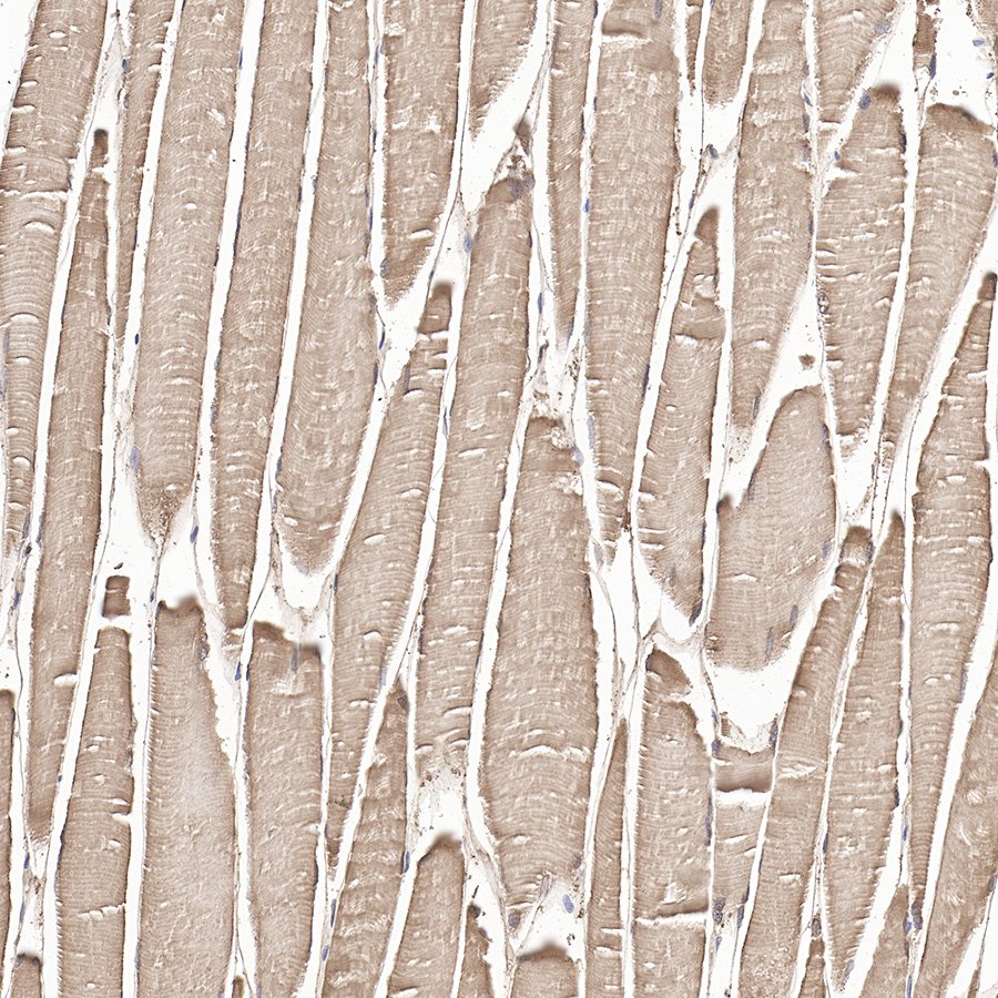
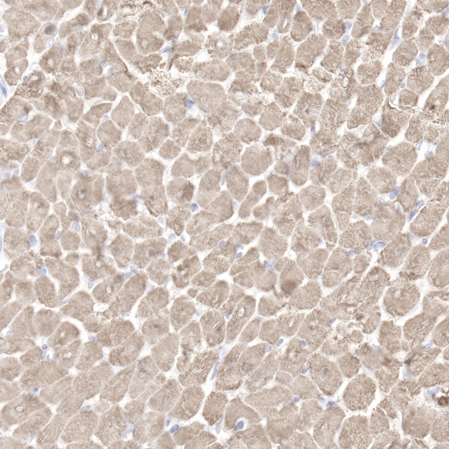
IHC shows positive staining in paraffin-embedded human skeletal muscle.
Anti-Myoglobin antibody was used at 1/1000 dilution, followed by a Goat Anti-Rabbit IgG H&L (HRP) ready to use. Counterstained with hematoxylin.
Heat mediated antigen retrieval with Tris/EDTA buffer pH9.0 was performed before commencing with IHC staining protocol.
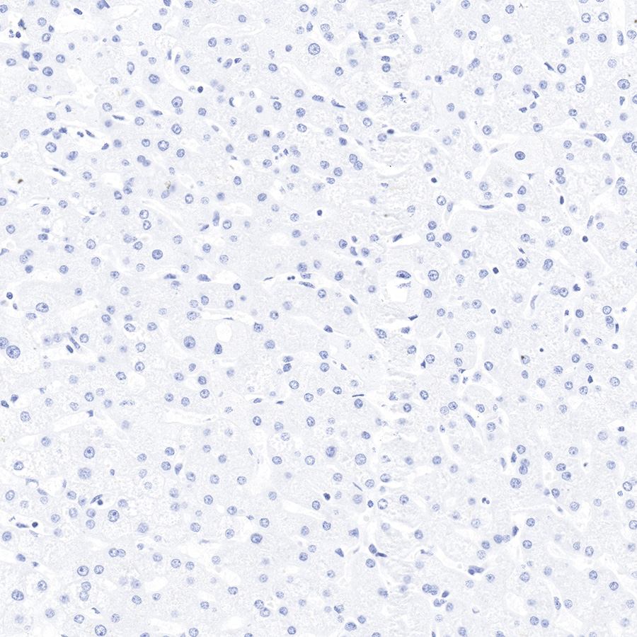
IHC shows negative staining in paraffin-embedded human liver.
Anti-Myoglobin antibody was used at 1/1000 dilution, followed by a Goat Anti-Rabbit IgG H&L (HRP) ready to use. Counterstained with hematoxylin.
Heat mediated antigen retrieval with Tris/EDTA buffer pH9.0 was performed before commencing with IHC staining protocol.
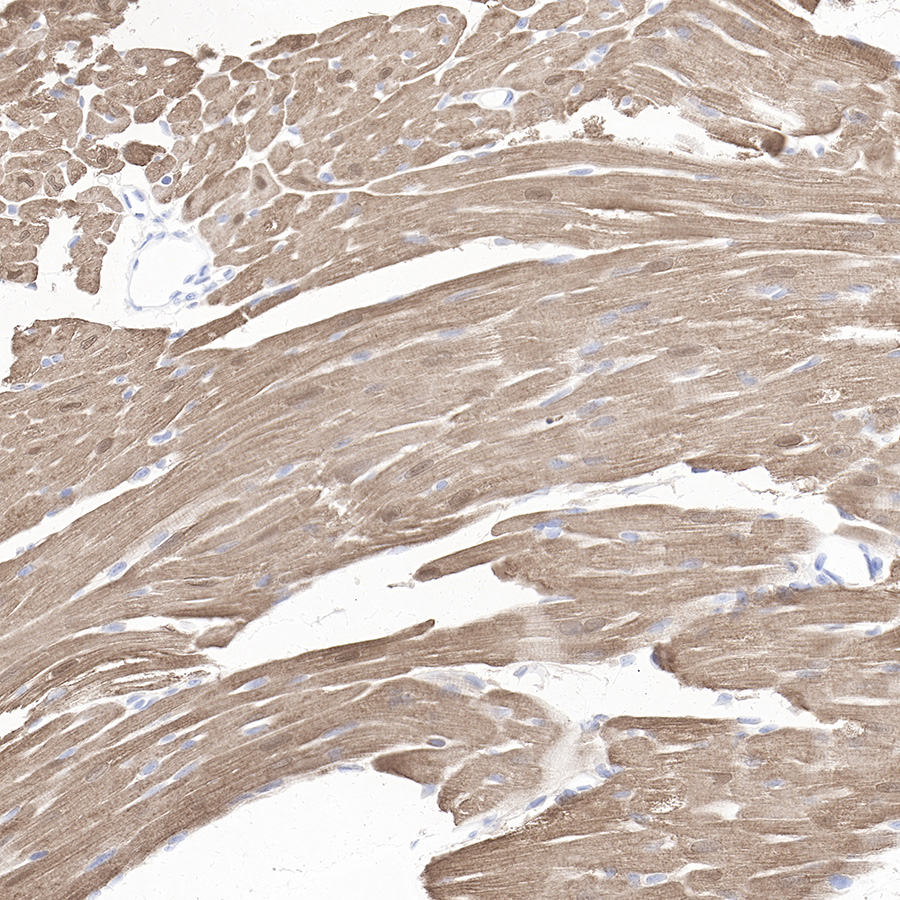
IHC shows positive staining in paraffin-embedded mouse heart.
Anti-Myoglobin antibody was used' at 1/1000 dilution, followed by a Goat Anti-Rabbit IgG H&L (HRP) ready to use. Counterstained with hematoxylin.
Heat mediated antigen retrieval with Tris/EDTA buffer pH9.0 was performed before commencing with IHC staining protocol.
IHC shows positive staining in paraffin-embedded rat heart.
Anti-Myoglobin antibody was used' at 1/1000 dilution, followed by a Goat Anti-Rabbit IgG H&L (HRP) ready to use. Counterstained with hematoxylin.
Heat mediated antigen retrieval with Tris/EDTA buffer pH9.0 was performed before commencing with IHC staining protocol.
免疫荧光
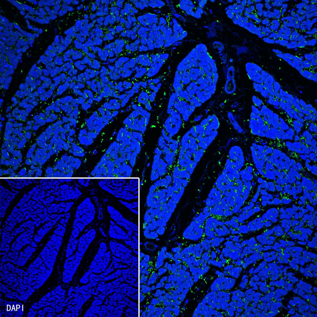
IF shows positive staining in paraffin-embedded human cardiac muscle. Anti-Myoglobin antibody was used at 1/1000 dilution (Green) and incubated overnight at 4°C. Goat polyclonal Antibody to Rabbit IgG - H&L (Alexa Fluor® 488) was used as secondary antibody at 1/1000 dilution. Counterstained with DAPI (Blue). Heat mediated antigen retrieval with Tris/EDTA buffer pH9.0 was performed before commencing with IF staining protocol.
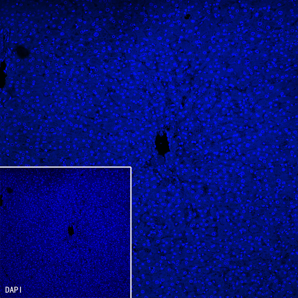
Negative control: IF shows positive staining in paraffin-embedded human liver. Anti-Myoglobin antibody was used at 1/1000 dilution and incubated overnight at 4°C. Goat polyclonal Antibody to Rabbit IgG - H&L (Alexa Fluor® 488) was used as secondary antibody at 1/1000 dilution. Counterstained with DAPI (Blue). Heat mediated antigen retrieval with Tris/EDTA buffer pH9.0 was performed before commencing with IF staining protocol.

评论(0)