


 大包装询价
大包装询价 产品介绍 评论(0)
宿主来源
Rabbit抗原名称
p53分子别名
Antigen NY-CO-13, Phosphoprotein p53, Tumor suppressor p53, TP53免疫原
Synthetic Peptide细胞定位
Cytoplasm, NucleusAccession
P04637克隆号
SDT-100-72抗体类型
Rabbit mAb应用
ICFCM, IHC-P, ICC, WB反应种属 ?
Hu纯化方式
Protein A浓度
1 mg/ml标记
Unconjugated性状
Liquid缓冲体系
PBS
储存条件
12 months from date of receipt, 4°C as supplied
| 应用 | 稀释度 |
|---|---|
| WB | 1:2500 |
| IHC-P | 1:1000 |
| ICC | 1:500 |
| ICFCM | 1:250 |
p53, also known as Tumor protein P53, cellular tumor antigen p53 (UniProt name), or transformation-related protein 53 (TRP53) is a regulatory protein that is often mutated in human cancers. The p53 proteins (originally thought to be, and often spoken of as, a single protein) are crucial in vertebrates, where they prevent cancer formation. As such, p53 has been described as "the guardian of the genome" because of its role in conserving stability by preventing genome mutation. Hence TP53 is classified as a tumor suppressor gene.
验证数据
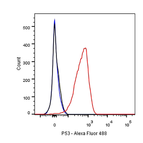
Flow cytometric analysis of HT-29 cells labelling p53 antibody at 1/250 dilution (0.1ug)/ (red) compared with a Rabbit monoclonal IgG (Black) isotype control and an unlabelled control (cells without incubation with primary antibody and secondary antibody) (Blue). Goat Anti-Rabbit IgG Alexa Fluor® 488 was used as the secondary antibody.
免疫印迹
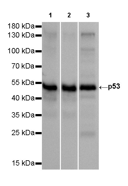
WB result of p53 Rabbit mAb
Primary antibody: p53 Rabbit mAb at 1/2500 dilution
Lane 1: A431 whole cell lysate 20 µg
Lane 2: HT-29 whole cell lysate 20 µg
Lane 3: T47D whole cell lysate 20 µgSecondary antibody: Goat Anti-Rabbit IgG, (H+L), HRP conjugated at 1/10000 dilution
Predicted MW: 53 kDa
Observed MW: 53 kDa
Exposure time: 10s
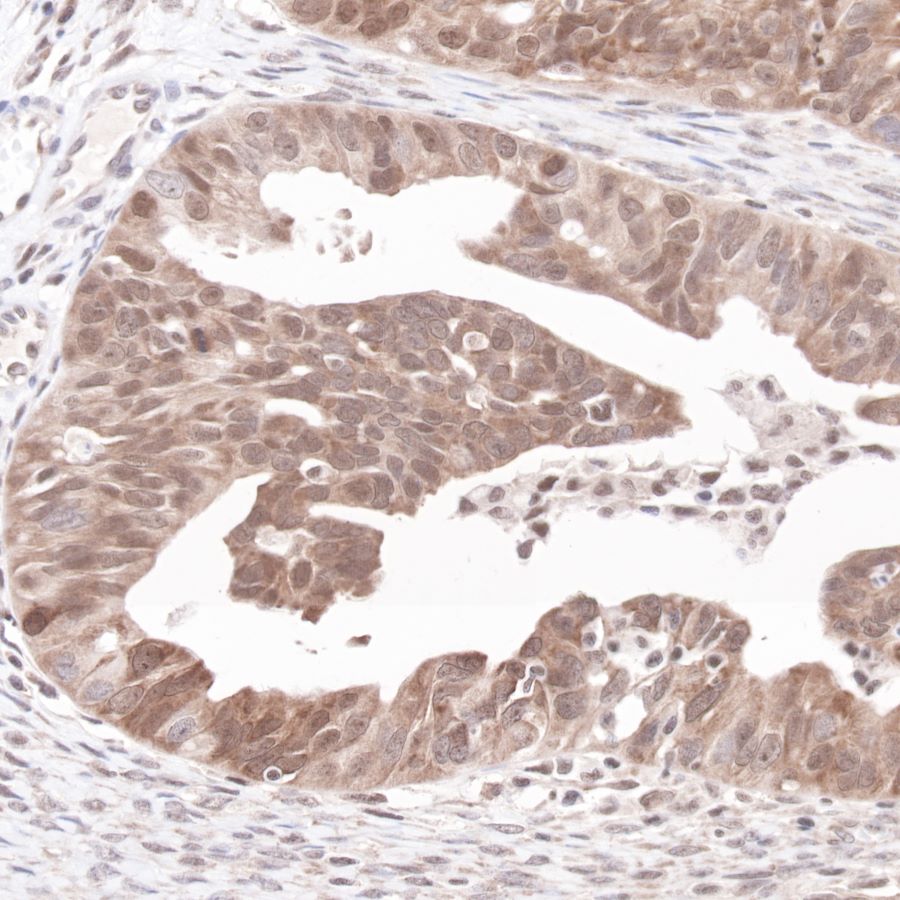
免疫组化
IHC shows positive staining in paraffin-embedded human colon cancer.
Anti-p53 antibody was used at 1/1000 dilution, followed by a Goat Anti-Rabbit IgG H&L (HRP) ready to use. Counterstained with hematoxylin.
Heat mediated antigen retrieval with Tris/EDTA buffer pH9.0 was performed before commencing with IHC staining protocol.
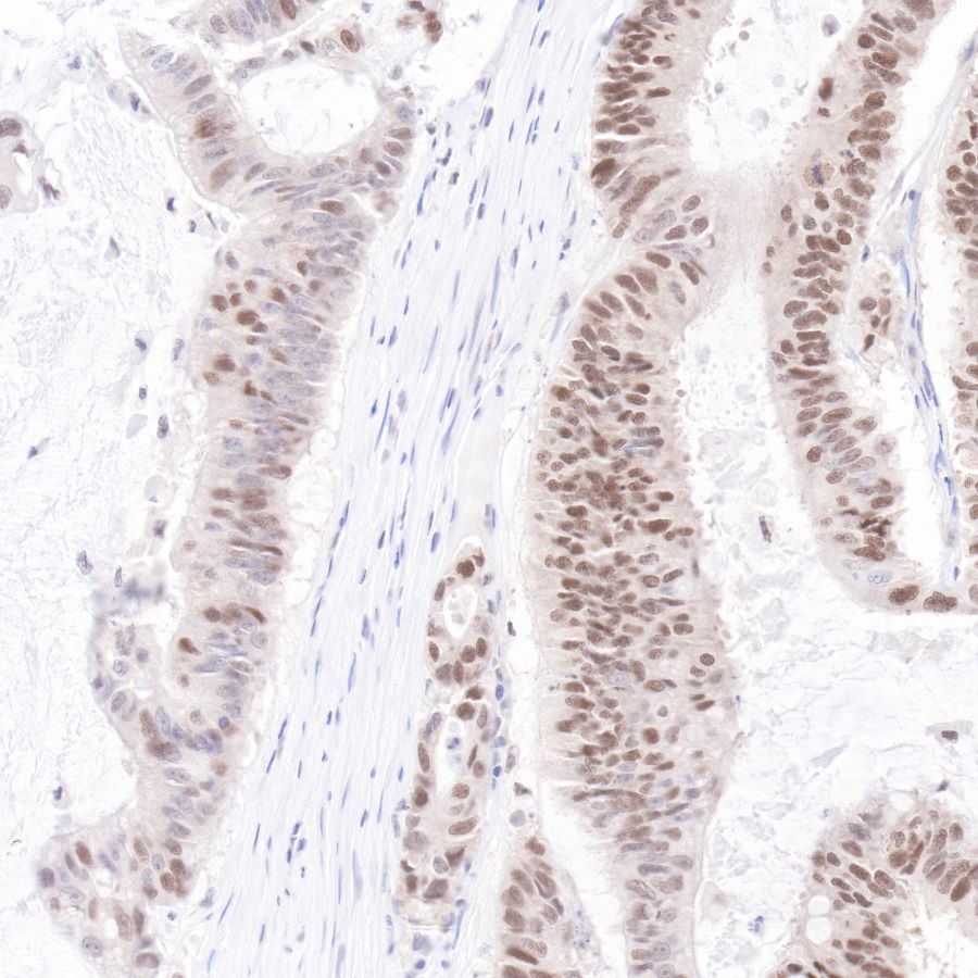
IHC shows positive staining in paraffin-embedded human colon cancer.
Anti-p53 antibody was used at 1/1000 dilution, followed by a Goat Anti-Rabbit IgG H&L (HRP) ready to use.
Counterstained with hematoxylin.
Heat mediated antigen retrieval with Tris/EDTA buffer pH9.0 was performed before commencing with IHC staining protocol.
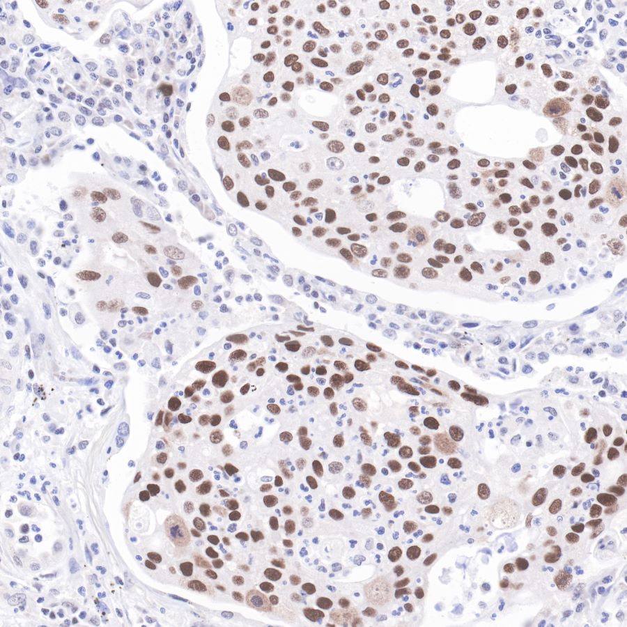
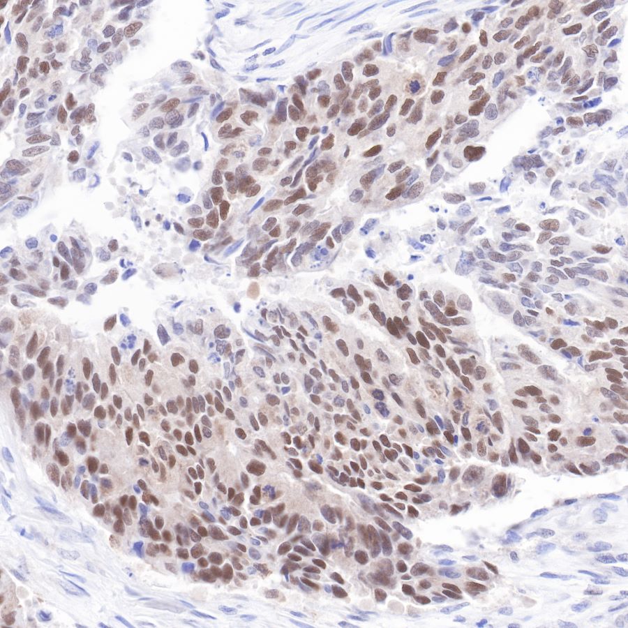
IHC shows positive staining in paraffin-embedded human lung cancer.
Anti-p53 antibody was used at 1/1000 dilution, followed by a Goat Anti-Rabbit IgG H&L (HRP) ready to use. Counterstained with hematoxylin.
Heat mediated antigen retrieval with Tris/EDTA buffer pH9.0 was performed before commencing with IHC staining protocol.
IHC shows positive staining in paraffin-embedded human ovarian cancer.
Anti-p53 antibody was used at 1/1000 dilution, followed by a Goat Anti-Rabbit IgG H&L (HRP) ready to use. Counterstained with hematoxylin.
Heat mediated antigen retrieval with Tris/EDTA buffer pH9.0 was performed before commencing with IHC staining protocol.
免疫细胞化学
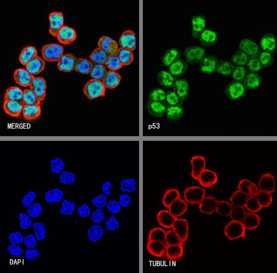
ICC shows positive staining in HT-29 cells. Anti-p53 antibody was used at 1/500 dilution (Green) and incubated overnight at 4°C. Goat polyclonal Antibody to Rabbit IgG - H&L (Alexa Fluor® 488) was used as secondary antibody at 1/1000 dilution. The cells were fixed with 4% PFA and permeabilized with 0.1% PBS-Triton X-100. Nuclei were counterstained with DAPI (Blue). Counterstain with tubulin (Red).

评论(0)