
产品介绍 评论(0)
宿主来源
Rabbit抗原名称
ErbB2分子别名
HER2, MLN19, NEU, NGL,CD340,ERBB2免疫原
Synthetic Peptide细胞定位
MembraneAccession
P04626克隆号
SDT-069-57抗体类型
Rabbit mAb应用
IHC-P ? ,ICC ? ,WB ,IF ?反应种属 ?
Hu纯化方式
Protein A浓度
0.5 mg/ml性状
Liquid缓冲体系
PBS, 40% Glycerol, 0.05%BSA, 0.03% Proclin 300
储存条件
12 months from date of receipt / reconstitution, -20 °C as supplied
| 应用 | 稀释度 |
|---|---|
| WB | 1:10000-1:50000 |
| IHC-P | 1:1000 |
| ICC | 1:500 |
| IF | 1:500 |
Receptor tyrosine-protein kinase erbB-2 is a protein that in humans is encoded by the ERBB2 gene. ERBB is abbreviated from erythroblastic oncogene B. The human protein is also frequently referred to as HER2 (human epidermal growth factor receptor 2) or CD340 (cluster of differentiation 340). HER2 is a member of the human epidermal growth factor receptor (HER/EGFR/ERBB) family. But contrary to other member of the ERBB family, HER2 does not directly bind ligand. HER2 activation results from heterodimerization with another ERBB member or by homodimerization when HER2 concentration are high, for instance in cancer. Amplification or over-expression of this oncogene has been shown to play an important role in the development and progression of certain aggressive types of breast cancer. In recent years the protein has become an important biomarker and target of therapy for approximately 30% of breast cancer patients.
免疫印迹

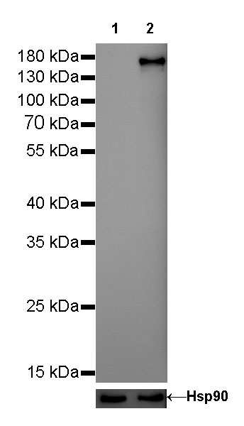
WB result of ErbB2 Rabbit mAb
Primary antibody: ErbB2 Rabbit mAb at 1/50000 dilution
Lane 1: MCF7 whle cell lysate 5 µg
Lane 2: SK-BR-3 whole cell lysate 5 µg
Low expression control: MCF7 whole cell lysateSecondary antibody: Goat Anti-Rabbit IgG, (H+L), HRP conjugated at 1/10000 dilution
Predicted MW: 185 kDa
Observed MW: 185 kDa
Exposure time: 30s
免疫组化

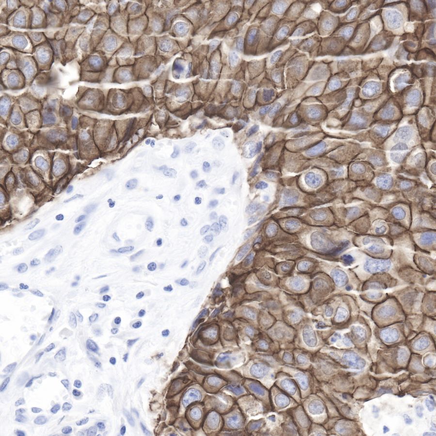
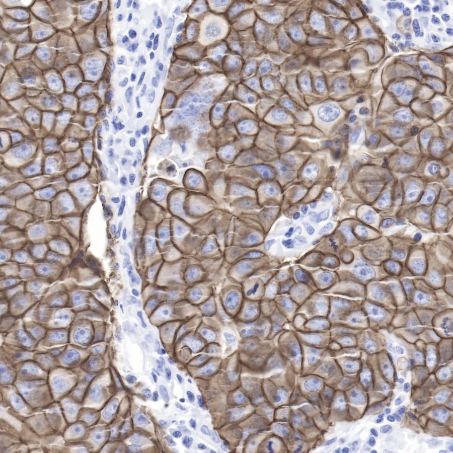
IHC shows positive staining in paraffin-embedded human breast cancer.
Anti-ErbB2 antibody was used at 1/1000 dilution, followed by a Goat Anti-Rabbit IgG H&L (HRP) ready to use.
Counterstained with hematoxylin.
Heat mediated antigen retrieval with Tris/EDTA buffer pH9.0 was performed before commencing with IHC staining protocol.

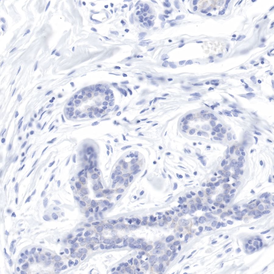
IHC shows negative staining in paraffin-embedded human breast. Anti-ErbB2 antibody was used at 1/1000 dilution, followed by a Goat Anti-Rabbit IgG H&L (HRP) ready to use.
Counterstained with hematoxylin.
Heat mediated antigen retrieval with Tris/EDTA buffer pH9.0 was performed before commencing with IHC staining protocol.
免疫细胞化学
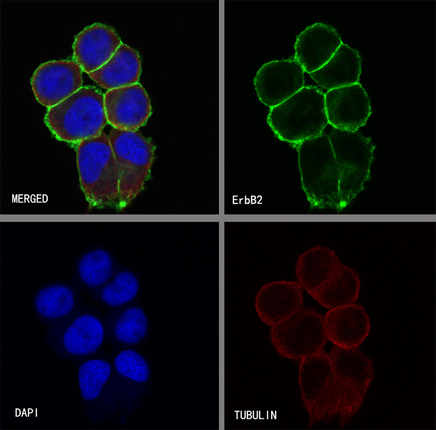
ICC shows positive staining in SK-BR-3 cells. Anti-ErbB2 antibody was used at 1/500 dilution (Green) and incubated overnight at 4°C. Goat polyclonal Antibody to Rabbit IgG - H&L (Alexa Fluor® 488) was used as secondary antibody at 1/1000 dilution. The cells were fixed with 100% ice-cold methanol and permeabilized with 0.1% PBS-Triton X-100. Nuclei were counterstained with DAPI (Blue). Counterstain with tubulin (red).
免疫荧光
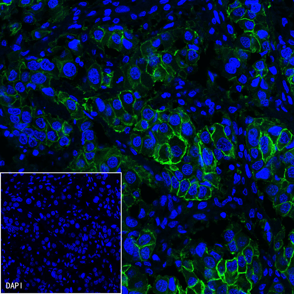
IF shows positive staining in paraffin-embedded human breast cancer. Anti-ErbB2 antibody was used at 1/500 dilution (Green) and incubated overnight at 4°C. Goat polyclonal Antibody to Rabbit IgG - H&L (Alexa Fluor® 488) was used as secondary antibody at 1/1000 dilution. Counterstained with DAPI (Blue). Heat mediated antigen retrieval with EDTA buffer pH9.0 was performed before commencing with IF staining protocol.
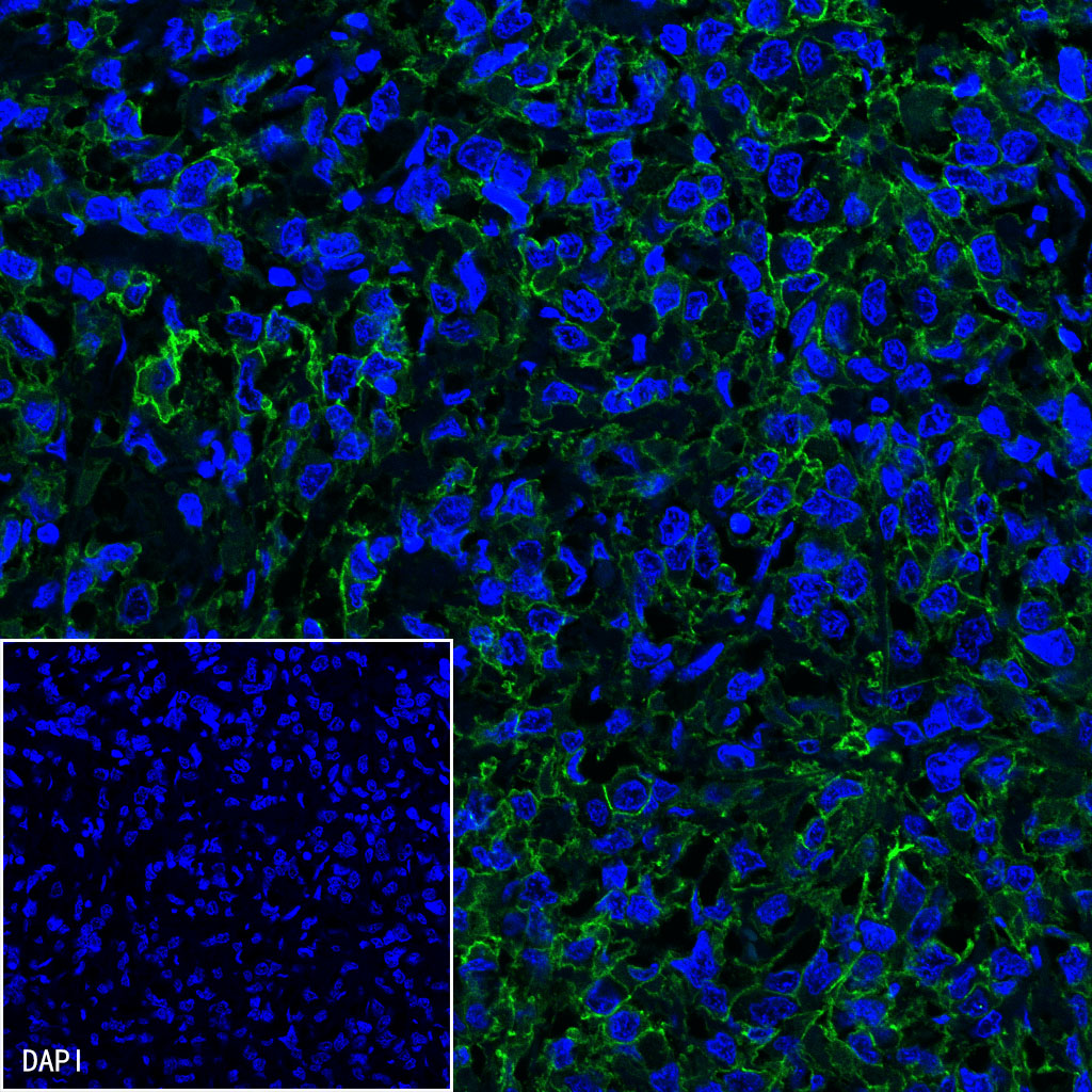
IF shows positive staining in paraffin-embedded human breast cancer. Anti-ErbB2 antibody was used at 1/500 dilution (Green) and incubated overnight at 4°C. Goat polyclonal Antibody to Rabbit IgG - H&L (Alexa Fluor® 488) was used as secondary antibody at 1/1000 dilution. Counterstained with DAPI (Blue). Heat mediated antigen retrieval with EDTA buffer pH9.0 was performed before commencing with IF staining protocol.
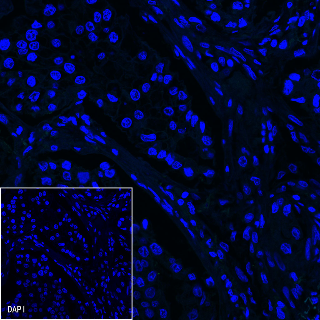
Negative control: IF shows negative staining in paraffin-embedded human lung adenocarcinoma. Anti-ErbB2 antibody was used at 1/500 dilution and incubated overnight at 4°C. Goat polyclonal Antibody to Rabbit IgG - H&L (Alexa Fluor® 488) was used as secondary antibody at 1/1000 dilution. Counterstained with DAPI (Blue). Heat mediated antigen retrieval with EDTA buffer pH9.0 was performed before commencing with IF staining protocol.


 申请试用
申请试用 
评论(0)