


 大包装询价
大包装询价 产品介绍 评论(0)
宿主来源
Rabbit抗原名称
Cytokeratin 7分子别名
Cytokeratin-7 (CK-7), Keratin-7 (K7), Sarcolectin, Type-II keratin Kb7, KRT7免疫原
N/A细胞定位
Cell membrane, IntracellularAccession
P08729克隆号
SDT-R028抗体类型
Rabbit mAb应用
ICFCM, IHC-P, ICC, WB, IP反应种属 ?
Hu纯化方式
Protein A浓度
1 mg/ml性状
Liquid缓冲体系
PBS储存条件
12 months from date of receipt, 4°C as supplied
| 应用 | 稀释度 |
|---|---|
| WB | 1:500 |
| IHC-P | 1:1000 |
| ICC | 1:50 |
| ICFCM | 1:500 |
| IP | 1: 25 |
Cytokeratin 7 is the intermediate silk protein mainly expressed in epithelial cells. The heterodine of keratin consists of a polymerization filament by an acid -keratin (or type I keratin, keratin 9 to 23) and alkaline keratin (or type II keratin, keratin 1 to 8). Expression similar to but more limited than keratins 8 and 18 in simple, pseudostratified and ductal epithelium, mesothelium and urothelium.It's enerally expressed (with some variation) in adenocarcinoma of lung, breast, thyroid, endometrium, cervix, ovary, salivary gland, upper GI tract, urothelial carcinoma, papillary renal cell carcinoma and Paget disease.It's generally negative (with some variation) in colorectal carcinoma, Merkel cell carcinoma, hepatocellular carcinoma, prostatic adenocarcinoma, adrenocortical tumors and squamous cell carcinoma.
免疫印迹

WB result of Cytokeratin 7 Rabbit mAb
Primary antibody: Cytokeratin 7 Rabbit mAb at 1/500 dilution
Lane 1: MCF7 whole cell lysate 20 µg
Lane 2: T47D whole cell lysate 20 µg
Lane 3: Hela whole cell lysate 20 µg
Lane 4: SK-OV-3 whole cell lysate 20 µg
Negative control: MCF7 whole cell lysateSecondary antibody: Goat Anti-Rabbit IgG, (H+L), HRP conjugated at 1/10000 dilution
Predicted MW: 52 kDa
Observed MW: 40~55 kDa
流式分析
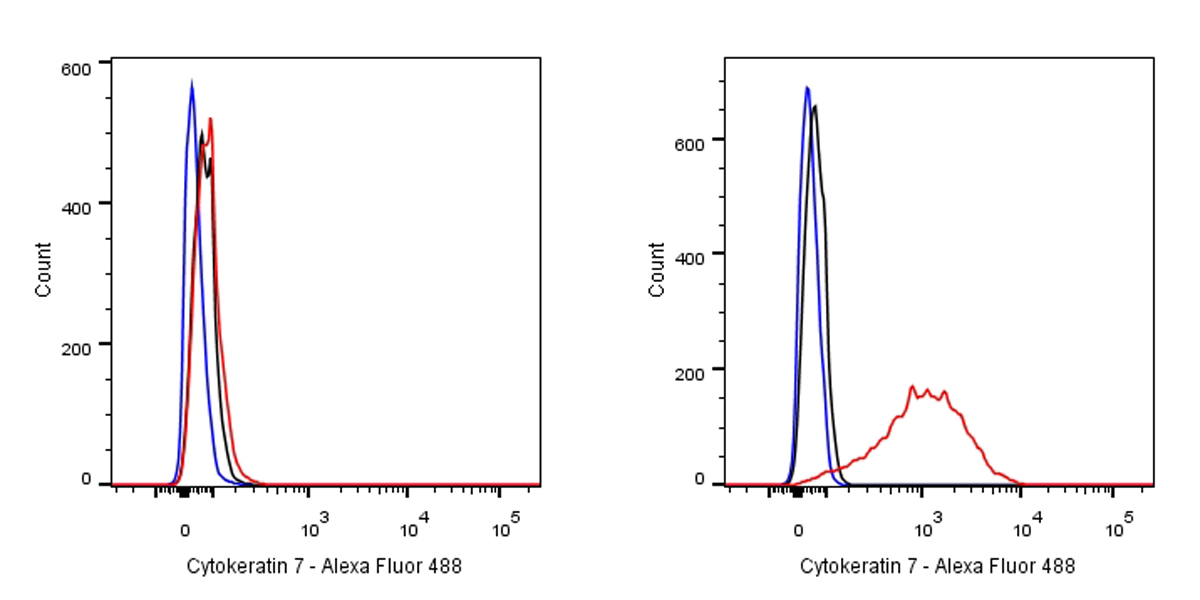
Flow cytometric analysis of MCF7 (left) / HeLa (right) cells labelling Cytokeratin-7 antibody at 1/500 (0.1ug) dilution/ (red) compared with a Rabbit monoclonal IgG (Black) isotype control and an unlabelled control (cells without incubation with primary antibody and secondary antibody) (Blue). Goat Anti-Rabbit IgG Alexa Fluor® 488 was used as the secondary antibody. Negative control: MCF7
免疫沉淀
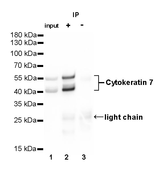
Cytokeratin 7 Rabbit mAb at 1/25 dilution (1µg) immunoprecipitating Cytokeratin 7 in 0.4mg T-47D whole cell lysate.
Western blot was performed on the immunoprecipitate using Cytokeratin 7 Rabbit mAb at 1/1000 dilution.
Secondary antibody (HRP) for IP was used at 1/400 dilution.
Lane 1 : T-47D whole cell lysate 20µg(input)
Lane 2 : Cytokeratin 7 Rabbit mAb IP in T-47D whole cell lysate
Lane 3 : Rabbit monoclonal IgG IP in T-47D whole cell lysate
Predicted MW: 52 kDa
Observed MW: 40-55 kDa
免疫组化
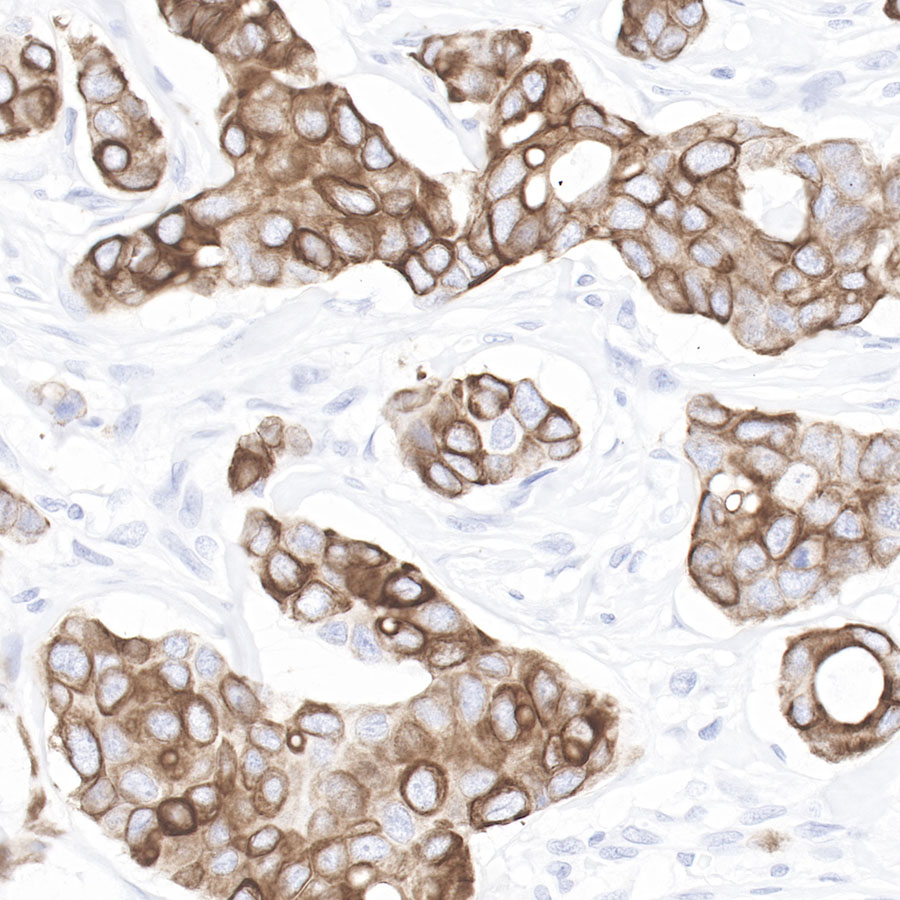
IHC shows positive staining in paraffin-embedded human breast cancer.
Anti-Cytokeratin 7 antibody was used at 1/1000 dilution, followed by a Goat Anti-Rabbit IgG H&L (HRP) ready to use.
Counterstained with hematoxylin.
Heat mediated antigen retrieval with Tris/EDTA buffer pH9.0 was performed before commencing with IHC staining protocol.
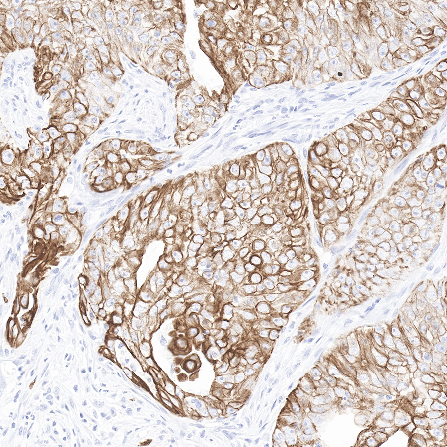
IHC shows positive staining in paraffin-embedded human endometrial cancer.
Anti-Cytokeratin 7 antibody was used at 1/1000 dilution, followed by a Goat Anti-Rabbit IgG H&L (HRP) ready to use.
Counterstained with hematoxylin.
Heat mediated antigen retrieval with Tris/EDTA buffer pH9.0 was performed before commencing with IHC staining protocol.
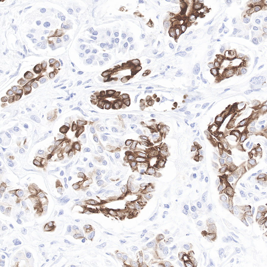
IHC shows positive staining in paraffin-embeddedhuman pancreas cancer.
Anti-Cytokeratin 7 antibody was used at 1/1000 dilution, followed by a Goat Anti-Rabbit IgG H&L (HRP) ready to use.
Counterstained with hematoxylin.
Heat mediated antigen retrieval with Tris/EDTA buffer pH9.0 was performed before commencing with IHC staining protocol.
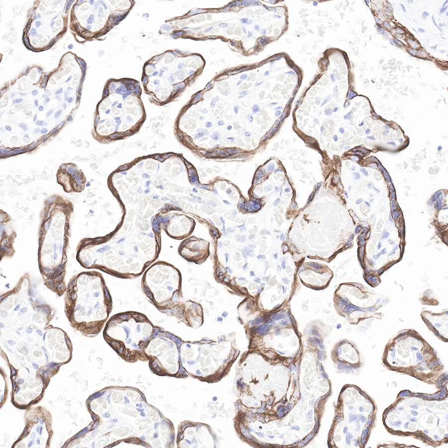
IHC shows positive staining in paraffin-embedded human placenta.
Anti-Cytokeratin 7 antibody was used at 1/1000 dilution, followed by a Goat Anti-Rabbit IgG H&L (HRP) ready to use.
Counterstained with hematoxylin.
Heat mediated antigen retrieval with Tris/EDTA buffer pH9.0 was performed before commencing with IHC staining protocol.
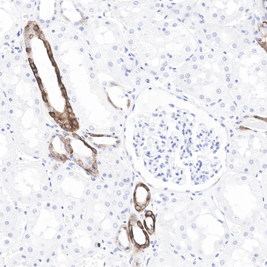
IHC shows positive staining in paraffin-embedded human kidney.
Anti-Cytokeratin 7 antibody was used at 1/1000 dilution, followed by a Goat Anti-Rabbit IgG H&L (HRP) ready to use. Counterstained with hematoxylin.
Heat mediated antigen retrieval with Tris/EDTA buffer pH9.0 was performed before commencing with IHC staining protocol.
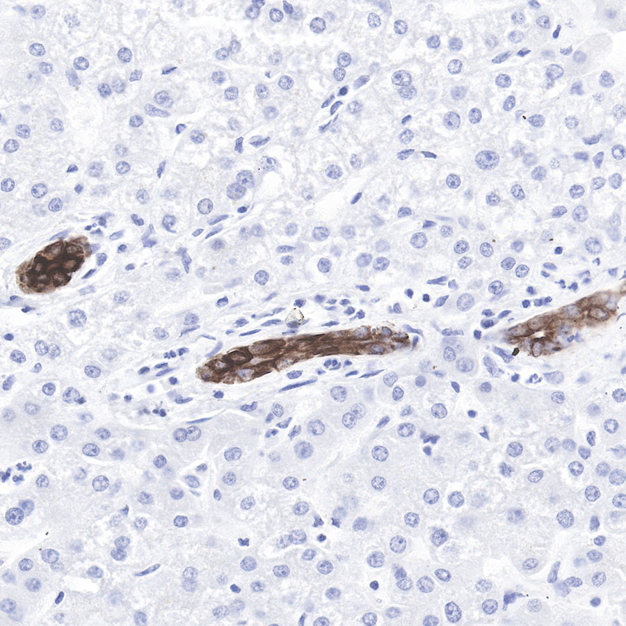
IHC shows positive staining in paraffin-embedded human liver.
Anti-Cytokeratin 7 antibody was used at 1/1000 dilution, followed by a Goat Anti-Rabbit IgG H&L (HRP) ready to use. Counterstained with hematoxylin.
Heat mediated antigen retrieval with Tris/EDTA buffer pH9.0 was performed before commencing with IHC staining protocol.
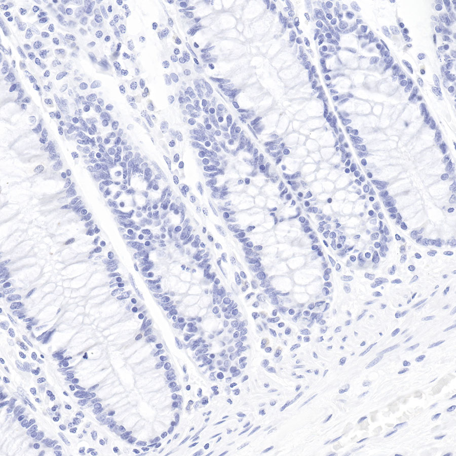
Negative control.IHC shows negative staining in paraffin-embedded human colon.
Anti-Cytokeratin 7 antibody was used at 1/500 dilution, followed by a Goat Anti-Rabbit IgG H&L (HRP) ready to use. Counterstained with hematoxylin.
Heat mediated antigen retrieval with Tris/EDTA buffer pH9.0 was performed before commencing with IHC staining protocol.

评论(0)