




 分享
分享产品介绍 评论(0)
宿主来源
Rabbit抗原名称
Calprotectin分子别名
Protein S100-A8, Calgranulin-A, MRP-8, S100 calcium-binding protein A8免疫原
Recombinant Protein细胞定位
SecretedAccession
P05109克隆号
SDT-049-44抗体类型
Rabbit mAb反应种属 ?
Hu, Ms, Rt纯化方式
Protein A浓度
0.1mg/ml性状
Liquid缓冲体系
PBS, 40% Glycerol, 0.05%BSA, 0.03% Proclin 300储存条件
12 months from date of receipt / reconstitution, -20 °C as supplied应用
IHC-P ?
WB
IP
稀释度
应用 稀释度 推荐种属 IHC-P 1:3200 Hu, Ms, Rt WB 1:200 Hu IP 1:25 Hu
- Calprotectin is a complex of the mammalian proteins S100A8 and S100A9. The proteins exist as homodimers but preferentially exist as S100A8/A9 heterodimers or heterotetramers (calprotectin) with antimicrobial, proinflammatory and prothrombotic properties. In the presence of calcium, calprotectin is capable of sequestering the transition metals iron, manganese and zinc via chelation. This metal sequestration affords the complex antimicrobial properties. Calprotectin is the only known antimicrobial manganese sequestration protein complex. Calprotectin comprises as much as 60% of the soluble protein content of the cytosol of a neutrophil, and it is secreted by an unknown mechanism during inflammation Faecal calprotectin has been used to detect intestinal inflammation (colitis or enteritis) and can serve as a biomarker for inflammatory bowel diseases. Blood based calprotectin (in serum and plasma) is used in diagnostics of multiple inflammatory diseases, including autoimmune diseases, like arthritis, and severe infections including sepsis.
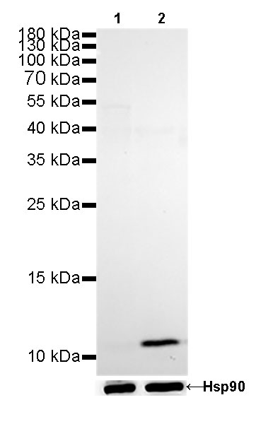
免疫印迹
WB result of Calprotectin Rabbit mAb
Primary antibody: Calprotectin Rabbit mAb at 1/200 dilution
Lane 1: C2C12 whole cell lysate 20 µg
Lane 2: THP-1 whole cell lysate 20 µg
Negative control: C2C12 whole cell lysateSecondary antibody: Goat Anti-Rabbit IgG, (H+L), HRP conjugated at 1/10000 dilution
Predicted MW: 11 kDa
Observed MW: 11 kDa
Exposure time: 180s
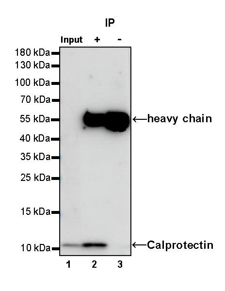
免疫沉淀
Calprotectin Rabbit mAb at 1/25 dilution (0.4 µg) immunoprecipitating Calprotectin in 0.4 mg THP-1 whole cell lysate.
Western blot was performed on the immunoprecipitate using Calprotectin Rabbit mAb at 1/1000 dilution.
Secondary antibody (HRP) for IP was used at 1/400 dilution.
Lane 1: THP-1 whole cell lysate 5 µg (Input)
Lane 2: Calprotectin Rabbit mAb IP in THP-1 whole cell lysate
Lane 3: Rabbit monoclonal IgG IP in THP-1 whole cell lysate
Predicted MW: 11 kDa
Observed MW: 11 kDa
Exposure time: 180 s(This blot was developed with high sensitivity substrate)
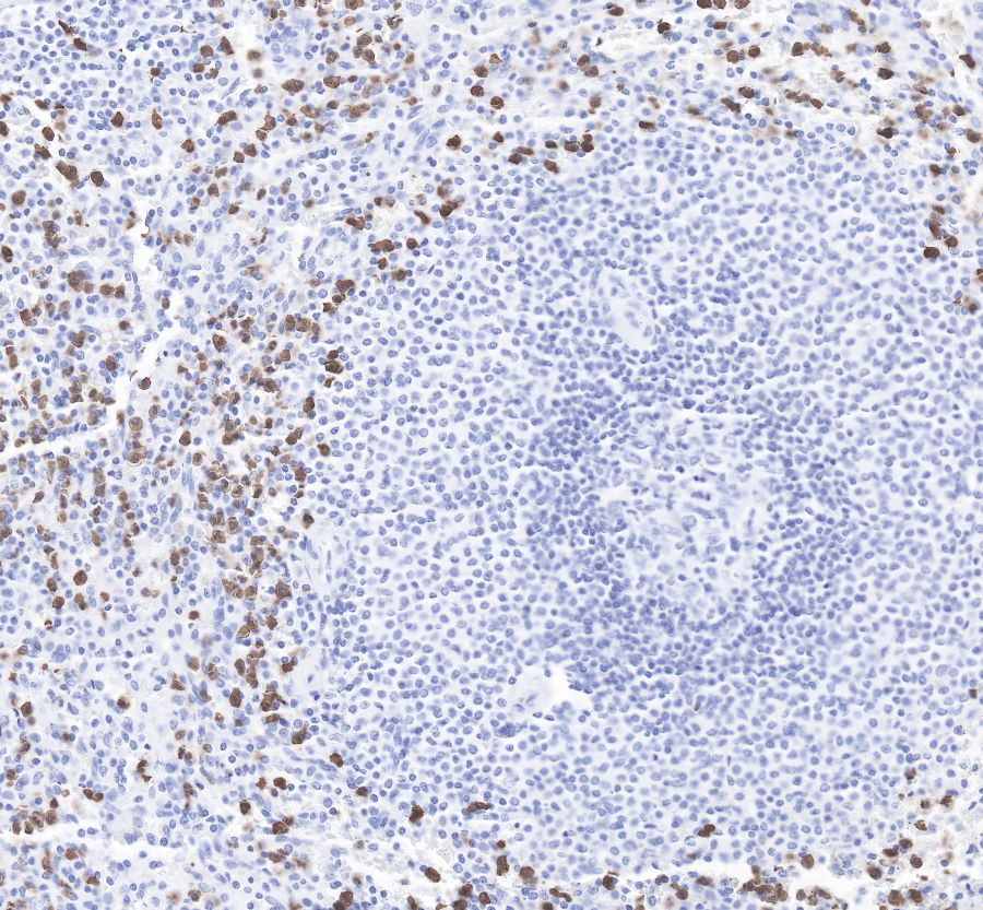
免疫组化
IHC shows positive staining in paraffin-embedded human spleen. Anti-Synaptophysin antibody was used at 1/3200 dilution, followed by a Goat Anti-Rabbit IgG H&L (HRP) ready to use.
Counterstained with hematoxylin.
Heat mediated antigen retrieval with Tris/EDTA buffer pH9.0 was performed before commencing with IHC staining protocol.
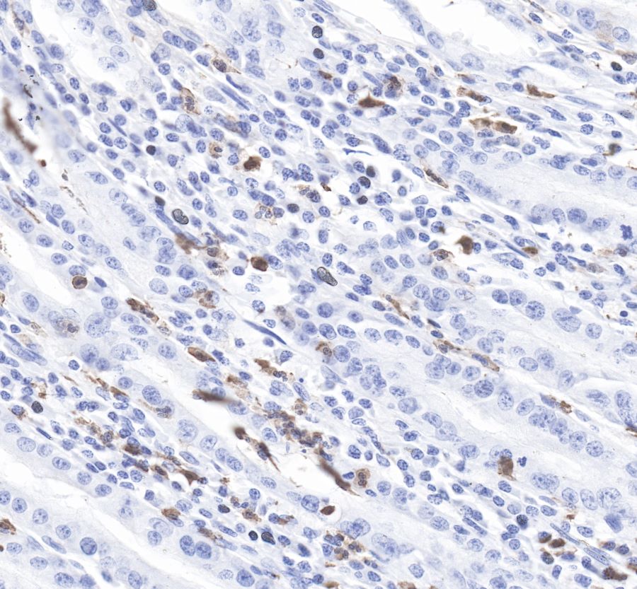
IHC shows positive staining in paraffin-embedded human stomach. Anti-Synaptophysin antibody was used at 1/3200 dilution, followed by a Goat Anti-Rabbit IgG H&L (HRP) ready to use.
Counterstained with hematoxylin.
Heat mediated antigen retrieval with Tris/EDTA buffer pH9.0 was performed before commencing with IHC staining protocol.
IHC shows positive staining in paraffin-embedded human liver.
Anti-Synaptophysin antibody was used at 1/3200 dilution, followed by a Goat Anti-Rabbit IgG H&L (HRP) ready to use.
Counterstained with hematoxylin.
Heat mediated antigen retrieval with Tris/EDTA buffer pH9.0 was performed before commencing with IHC staining protocol.
IHC shows positive staining in paraffin-embedded human cervix cancer.
Anti-Synaptophysin antibody was used at 1/3200 dilution, followed by a Goat Anti-Rabbit IgG H&L (HRP) ready to use.
Counterstained with hematoxylin.
Heat mediated antigen retrieval with Tris/EDTA buffer pH9.0 was performed before commencing with IHC staining protocol.
IHC shows positive staining in paraffin-embedded human lung cancer.
Anti-Synaptophysin antibody was used at 1/3200 dilution, followed by a Goat Anti-Rabbit IgG H&L (HRP) ready to use.
Counterstained with hematoxylin.
Heat mediated antigen retrieval with Tris/EDTA buffer pH9.0 was performed before commencing with IHC staining protocol.
IHC shows positive staining in paraffin-embedded mouse spleen. Anti-Synaptophysin antibody was used at 1/3200 dilution, followed by a Goat Anti-Rabbit IgG H&L (HRP) ready to use.
Counterstained with hematoxylin.
Heat mediated antigen retrieval with Tris/EDTA buffer pH9.0 was performed before commencing with IHC staining protocol.
IHC shows positive staining in paraffin-embedded rat spleen.
Anti-Synaptophysin antibody was used at 1/3200 dilution, followed by a Goat Anti-Rabbit IgG H&L (HRP) ready to use.
Counterstained with hematoxylin. Heat mediated antigen retrieval with Tris/EDTA buffer pH9.0 was performed before commencing with IHC staining protocol.

评论(0)