




 分享
分享产品介绍 评论(0)
宿主来源
Rabbit抗原名称
Synaptophysin分子别名
Major synaptic vesicle protein p38,SYP免疫原
Synthetic Peptide细胞定位
Intracellular, SynaptosomeAccession
P08247克隆号
SDT-137-21抗体类型
Rabbit mAb反应种属 ?
Hu, Ms, Rt预测反应种属
(反应种属缩写表)Bv, Sq纯化方式
Protein A浓度
0.25mg/ml性状
Liquid缓冲体系
PBS, 40% Glycerol, 0.05%BSA, 0.03% Proclin 300
储存条件
12 months from date of receipt / reconstitution, -20 °C as supplied
应用
ICFCM
IHC-P ?
ICC ?
WB
稀释度
应用 稀释度 推荐种属 WB 1:1000-1:10000 Ms, Rt IHC-P 1:4000(Ms) Ms IHC-P 1:1000(Hu) Ms ICFCM 1:500 Rt ICC 1:250 Rt
Synaptophysin, also known as the major synaptic vesicle protein p38, is a protein that in humans is encoded by the SYP gene. The protein is a synaptic vesicle glycoprotein with four transmembrane domains weighing 38kDa. It is present in neuroendocrine cells and in virtually all neurons in the brain and spinal cord that participate in synaptic transmission. It acts as a marker for neuroendocrine tumors, and its ubiquity at the synapse has led to the use of synaptophysin immunostaining for quantification of synapses.
免疫印迹
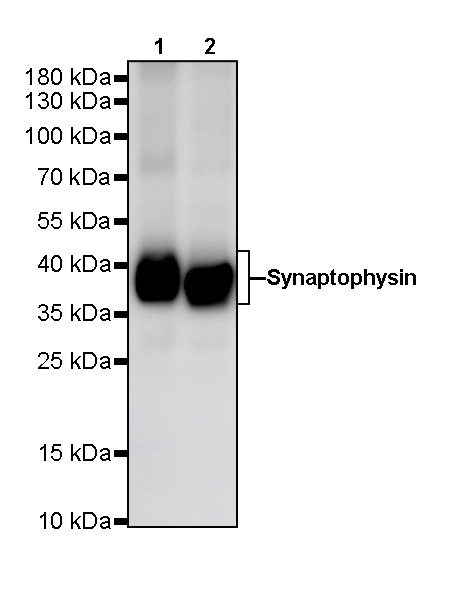
WB result of Synaptophysin Rabbit mAb
Primary antibody: Synaptophysin Rabbit mAb at 1/10000 dilution
Lane 1: mouse cerebellum lysate 20 µg
Lane 2: mouse brain lysate 20 µg
Secondary antibody: Goat Anti-Rabbit IgG, (H+L), HRP conjugated at 1/10000 dilution
Predicted MW: 38 kDa
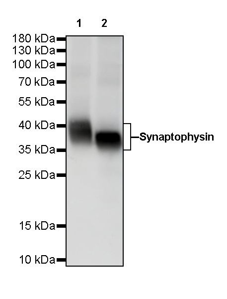
Observed MW: 38~42 kDaWB result of Synaptophysin Rabbit mAb
Primary antibody: Synaptophysin Rabbit mAb at 1/10000 dilution
Lane 1: PC-12 whole cell lysate 20 µg
Lane 2: rat brain lysate 20 µg
Secondary antibody: Goat Anti-Rabbit IgG, (H+L), HRP conjugated at 1/10000 dilution
Predicted MW: 38 kDa
Observed MW: 38~42 kDa
流式分析
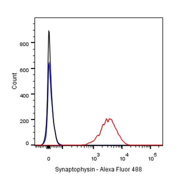
Flow cytometric analysis of PC-12 cells labelling Synaptophysin antibody at 1/500 (0.1ug) dilution/ (red) compared with a Rabbit monoclonal IgG (Black) isotype control and an unlabelled control (cells without incubation with primary antibody and secondary antibody) (Blue). Goat Anti-Rabbit IgG Alexa Fluor® 488 was used as the secondary antibody.
免疫组化
IHC shows positive staining in paraffin-embedded human pancreas.
Anti-Synaptophysin antibody was used at 1/1000 dilution, followed by a Goat Anti-Rabbit IgG H&L (HRP) ready to use.
Counterstained with hematoxylin.
Heat mediated antigen retrieval with Tris/EDTA buffer pH9.0 was performed before commencing with IHC staining protocol.
IHC shows positive staining in paraffin-embedded human brain.
Anti-Synaptophysin antibody was used at 1/1000 dilution, followed by a Goat Anti-Rabbit IgG H&L (HRP) ready to use.
Counterstained with hematoxylin.
Heat mediated antigen retrieval with Tris/EDTA buffer pH9.0 was performed before commencing with IHC staining protocol.
IHC shows positive staining in paraffin-embedded human colon.
Anti-Synaptophysin antibody was used at 1/1000 dilution, followed by a Goat Anti-Rabbit IgG H&L (HRP) ready to use.
Counterstained with hematoxylin.
Heat mediated antigen retrieval with Tris/EDTA buffer pH9.0 was performed before commencing with IHC staining protocol.
IHC shows positive staining in paraffin-embedded human medullary thyroid carcinoma.
Anti-Synaptophysin antibody was used at 1/1000 dilution, followed by a Goat Anti-Rabbit IgG H&L (HRP) ready to use.
Counterstained with hematoxylin.
Heat mediated antigen retrieval with Tris/EDTA buffer pH9.0 was performed before commencing with IHC staining protocol.
IHC shows positive staining in paraffin-embedded human small cell lung carcinoma.
Anti-Synaptophysin antibody was used at 1/1000 dilution, followed by a Goat Anti-Rabbit IgG H&L (HRP) ready to use.
Counterstained with hematoxylin.
Heat mediated antigen retrieval with Tris/EDTA buffer pH9.0 was performed before commencing with IHC staining protocol.
Negative tissue: IHC shows negative staining in paraffin-embedded human cervix cancer.
Anti-Synaptophysin antibody was used at 1/1000 dilution, followed by a Goat Anti-Rabbit IgG H&L (HRP) ready to use.
Counterstained with hematoxylin.
Heat mediated antigen retrieval with Tris/EDTA buffer pH9.0 was performed before commencing with IHC staining protocol.
IHC shows positive staining in paraffin-embedded mouse brain.
Anti-Synaptophysin antibody was used at 1/4000 dilution, followed by a Goat Anti-Rabbit IgG H&L (HRP) ready to use.
Counterstained with hematoxylin.
Heat mediated antigen retrieval with Tris/EDTA buffer pH9.0 was performed before commencing with IHC staining protocol.
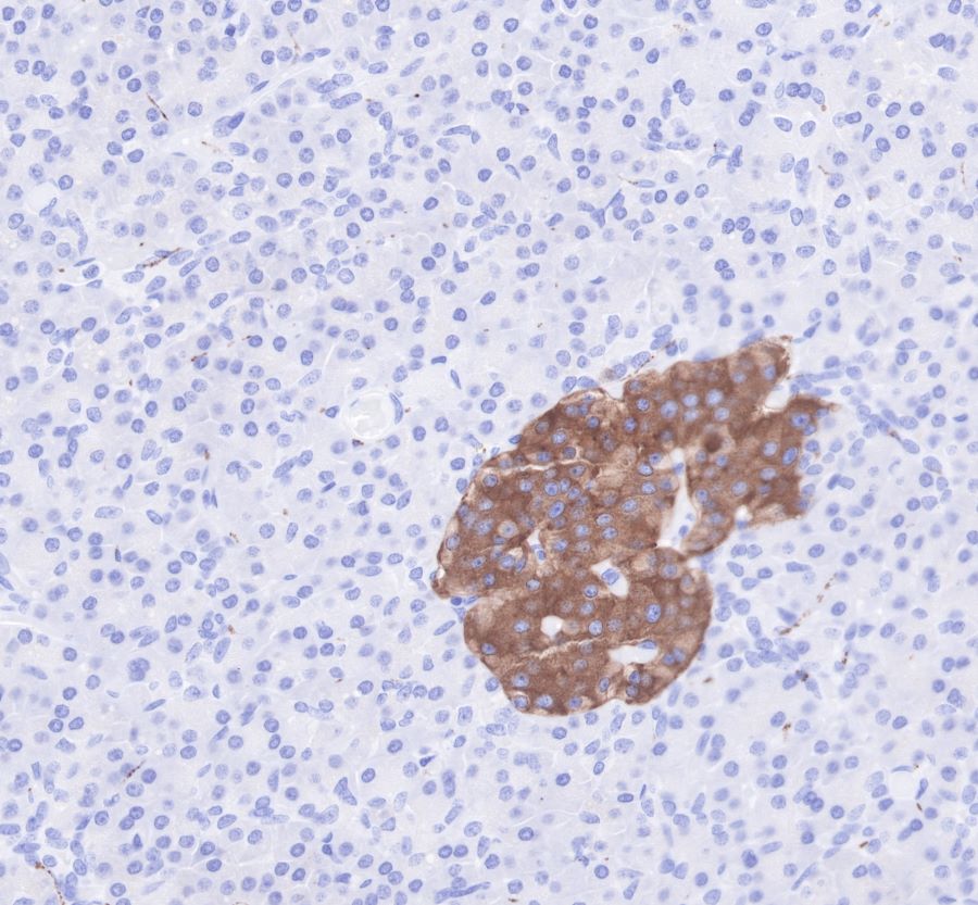
IHC shows positive staining in paraffin-embedded mouse pancreas.
Anti-Synaptophysin antibody was used at 1/4000 dilution, followed by a Goat Anti-Rabbit IgG H&L (HRP) ready to use.
Counterstained with hematoxylin.
Heat mediated antigen retrieval with Tris/EDTA buffer pH9.0 was performed before commencing with IHC staining protocol.
IHC shows positive staining in paraffin-embedded rat brain.
Anti-Synaptophysin antibody was used at 1/4000 dilution, followed by a Goat Anti-Rabbit IgG H&L (HRP) ready to use.
Counterstained with hematoxylin.
Heat mediated antigen retrieval with Tris/EDTA buffer pH9.0 was performed before commencing with IHC staining protocol.
IHC shows positive staining in paraffin-embedded rat pancreas.
Anti-Synaptophysin antibody was used at 1/4000 dilution, followed by a Goat Anti-Rabbit IgG H&L (HRP) ready to use.
Counterstained with hematoxylin.
Heat mediated antigen retrieval with Tris/EDTA buffer pH9.0 was performed before commencing with IHC staining protocol.
免疫细胞化学
ICC shows positive staining in PC-12 cells. Anti-Synatophysin antibody was used at 1/500 dilution (Green) and incubated overnight at 4°C. Goat polyclonal Antibody to Rabbit IgG - H&L (Alexa Fluor® 488) was used as secondary antibody at 1/1000 dilution. The cells were fixed with 4%PFA and permeabilized with 0.1% PBS-Triton X-100. Nuclei were counterstained with DAPI (Blue).Counterstain with tubulin (Red).

评论(0)