




 分享
分享产品介绍 评论(0)
宿主来源
Rabbit抗原名称
TCL1分子别名
Oncogene TCL-1, Protein p14 TCL1免疫原
Synthetic Peptide细胞定位
Cytoplasm, NucleusAccession
P56279克隆号
SDT-052-12抗体类型
Rabbit mAb反应种属 ?
Hu纯化方式
Protein A浓度
0.5mg/ml标记
Unconjugated性状
Liquid缓冲体系
PBS, 40% Glycerol, 0.05%BSA, 0.03% Proclin 300储存条件
12 months from date of receipt / reconstitution, -20 °C as supplied应用
IHC-P ?
WB
IP
稀释度
应用 稀释度 IHC-P 1:2000 WB 1:1000 IP 1:25
T-cell leukemia/lymphoma protein 1A is a protein that in humans is encoded by the TCL1A gene. The TCL1 locus on human chromosome 14q32.1 is activated in T-cell leukemias by translocations and inversions that juxtapose it to regulatory elements of T-cell receptor genes. TCL1A has been shown to interact with AKT1 and AKT2. Protein expression in immature T and B lymphoid cells and overexpression of TCL1 is considered critical in the oncogenetic transformation.
免疫印迹
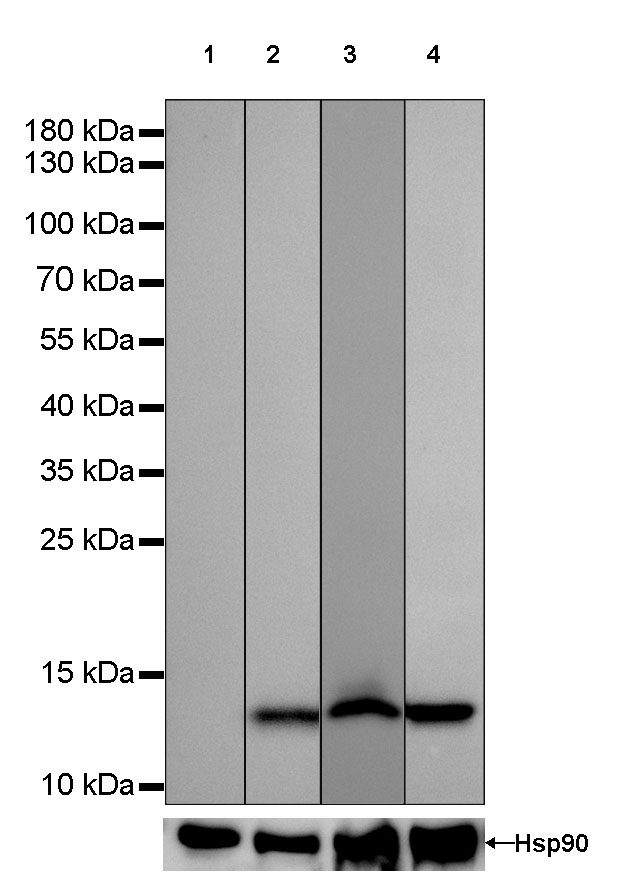
WB result of TCL1 Rabbit mAb
Primary antibody: TCL1 Rabbit mAb at 1/1000 dilution
Lane 1: HL-60 whole cell lysate 20 µg
Lane 2: Raji whole cell lysate 20 µg
Lane 3: Ramos whole cell lysate 20 µg
Lane 4: Daudi whole cell lysate 20 µg
Negative control: HL-60 whole cell lysateSecondary antibody: Goat Anti-Rabbit IgG, (H+L), HRP conjugated at 1/10000 dilution
Predicted MW: 13 kDa
Observed MW: 13 kDa
Exposure time: 6s
免疫沉淀
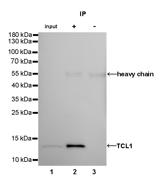
TCL1 Rabbit mAb at 1/25 dilution (2µg) immunoprecipitating TCL1 in 0.4mg Daudi whole cell lysate. Western blot was performed on the immunoprecipitate using TCL1 Rabbit mAb at 1/1000 dilution. Secondary antibody (HRP) for IP was used at 1/400 dilution.
Lane 1: Daudi whole cell lysate 10µg (input)
Lane 2: TCL1 Rabbit mAb IP in Daudi whole cell lysate
Lane 3: Rabbit monoclonal IgG IP in Daudi whole cell lysate
Predicted MW: 13 kDa
Observed MW: 13 kDa
Exposure time: 72s
免疫组化
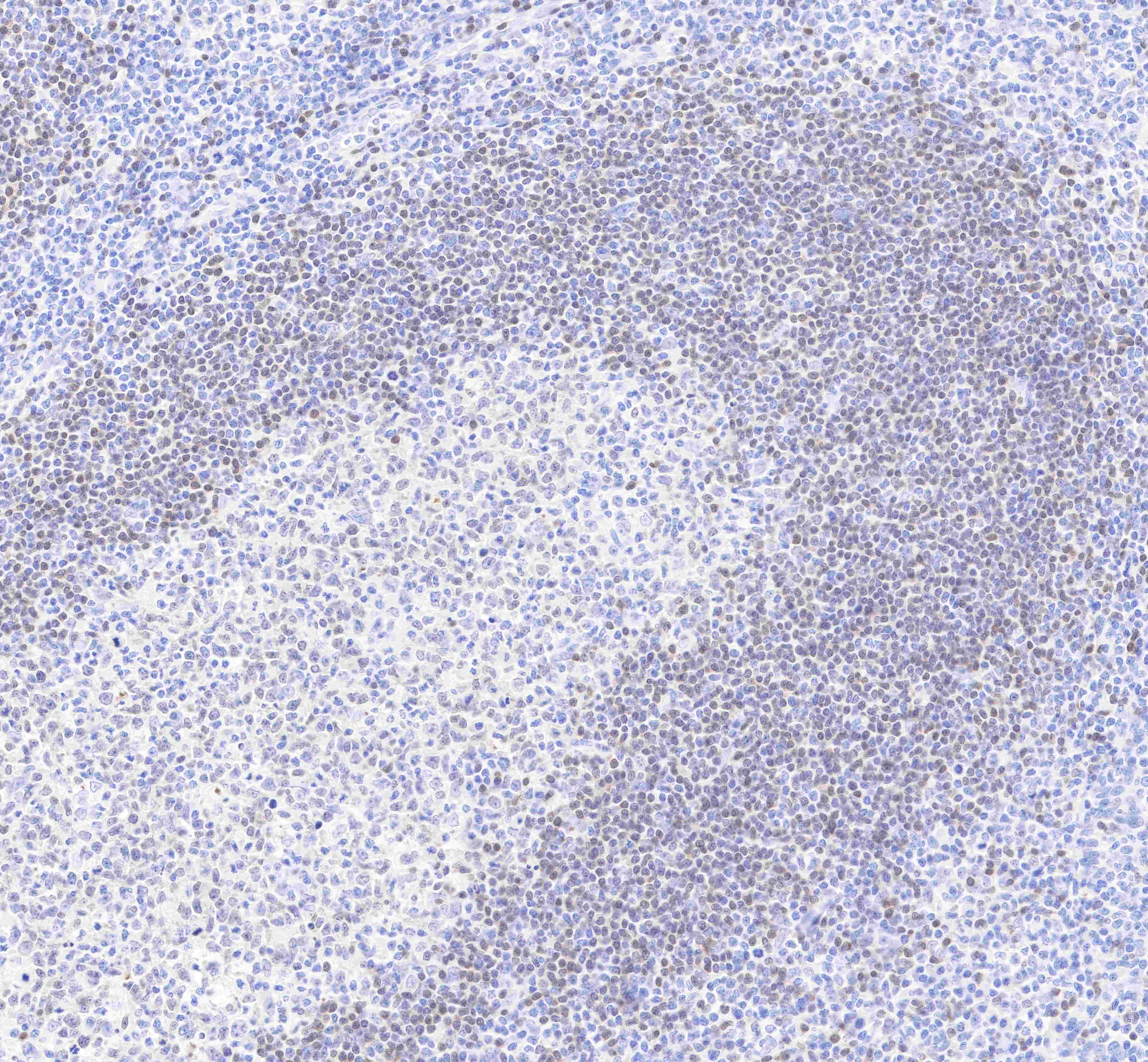
IHC shows positive staining in paraffin-embedded human tonsil.
Anti-TCL1 antibody was used at 1/2000 dilution, followed by a Goat Anti-Rabbit IgG H&L (HRP) ready to use. Counterstained with hematoxylin.
Heat mediated antigen retrieval with Tris/EDTA buffer pH9.0 was performed before commencing with IHC staining protocol.
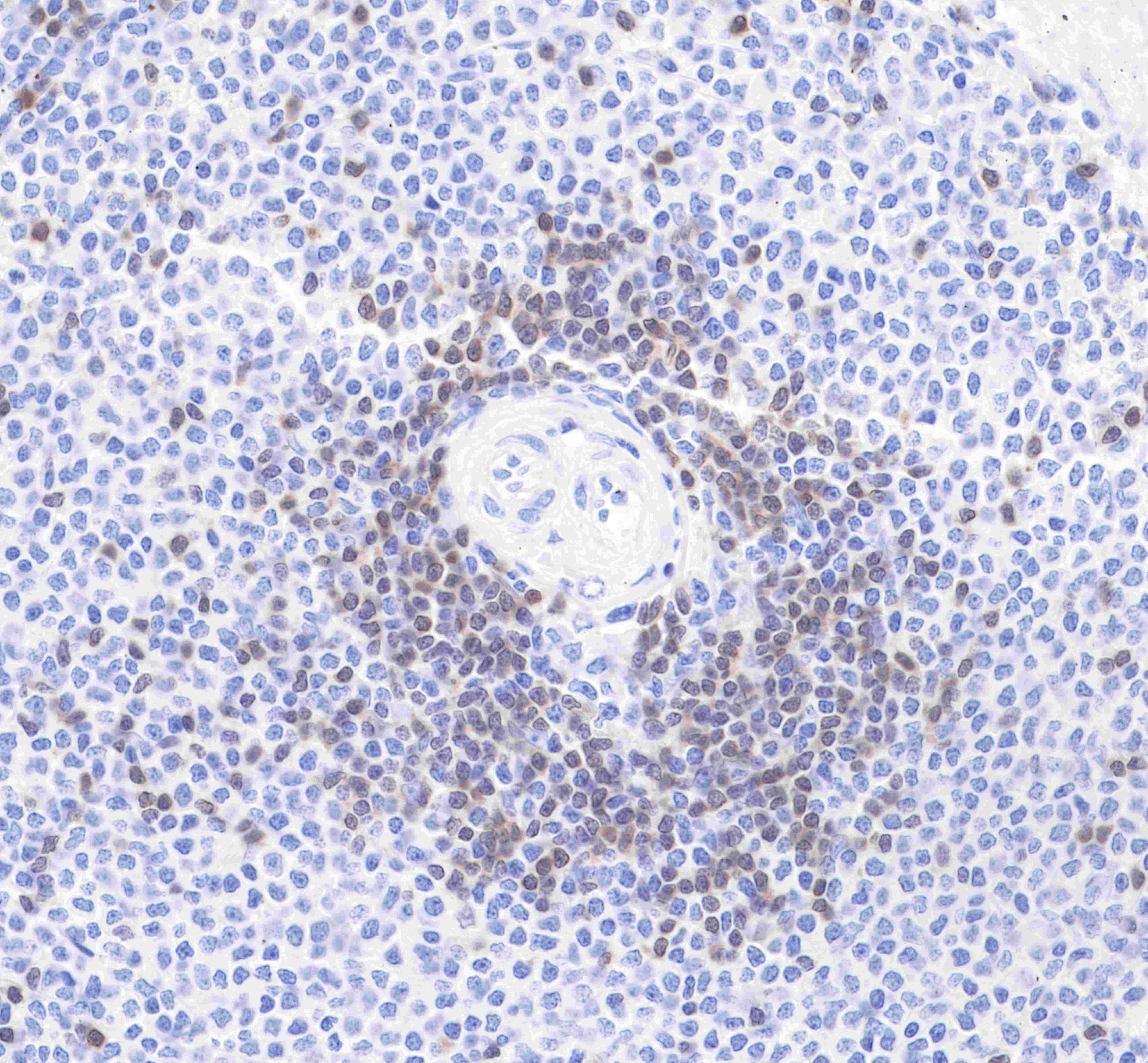
IHC shows positive staining in paraffin-embedded human spleen.
Anti-TCL1 antibody was used at 1/2000 dilution, followed by a Goat Anti-Rabbit IgG H&L (HRP) ready to use. Counterstained with hematoxylin.
Heat mediated antigen retrieval with Tris/EDTA buffer pH9.0 was performed before commencing with IHC staining protocol.

评论(0)