


 申请试用
申请试用 S-RMab® CD38 Recombinant Rabbit mAb (SDT-031-45-2) STAR
2'-phospho-ADP-ribosyl cyclase,2'-phospho-ADP-ribosyl cyclase/2'-phospho-cyclic-ADP-ribose transferase,2'-phospho-cyclic-ADP-ribose transferase,ADP-ribosyl cyclase 1 (ADPRC 1),Cyclic ADP-ribose hydrolase 1 (cADPr hydrolase 1),T10
产品介绍 评论(0)
宿主来源
Rabbit抗原名称
CD38分子别名
2'-phospho-ADP-ribosyl cyclase, 2'-phospho-ADP-ribosyl cyclase/2'-phospho-cyclic-ADP-ribose transferase,2'-phospho-cyclic-ADP-ribose transferase,ADP-ribosyl cyclase 1 (ADPRC 1),Cyclic ADP-ribose hydrolase 1 (cADPr hydrolase 1),T10免疫原
Synthetic Peptide细胞定位
MembraneAccession
P28907克隆号
SDT-031-45-2抗体类型
Rabbit mAb反应种属 ?
Hu纯化方式
Protein A浓度
0.05mg/ml标记
Unconjugated性状
Liquid缓冲体系
PBS, 40% Glycerol, 0.05%BSA, 0.03% Proclin 300储存条件
12 months from date of receipt / reconstitution, -20 °C as supplied
应用
稀释度
应用 稀释度 FCM 1:50 IHC-P 1: 500-1:1000 WB 1:500 ICC 1:50
CD38 (cluster of differentiation 38), also known as cyclic ADP ribose hydrolase is a glycoprotein found on the surface of many immune cells (white blood cells), including CD4+, CD8+, B lymphocytes and natural killer cells. CD38 also functions in cell adhesion, signal transduction and calcium signaling. The loss of CD38 function is associated with impaired immune responses, metabolic disturbances, and behavioral modifications including social amnesia possibly related to autism. The CD38 protein is a marker of cell activation. It has been connected to HIV infection, leukemias, myelomas, solid tumors, type II diabetes mellitus and bone metabolism, as well as some genetically determined conditions.
验证数据
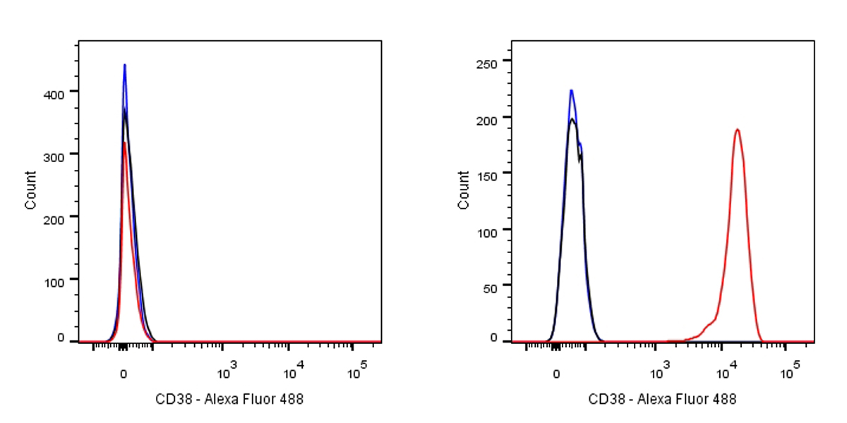
Flow cytometric analysis of HeLa (left) / Ramos (right) cells labelling CD38 antibody at 1/50 dilution (0.1ug)/ (red) compared with a Rabbit monoclonal IgG (Black) isotype control and an unlabelled control (cells without incubation with primary antibody and secondary antibody) (Blue). Goat Anti-Rabbit IgG Alexa Fluor® 488 at 1/1000 dilution was used as the secondary antibody. Negative control: HeLa
免疫印迹
WB result of CD38 Rabbit mAb
Primary antibody : CD38 Rabbit mAb at 1/500 dilution
Lane 1: HepG2 whole cell lysate 20 µg
Lane 2: Daudi whole cell stomach lysate 20 µg
Lane 3: Jurkat whole cell lysate 20 µg
Lane 4: Raji whole cell heart lysate 20 µg
Lane 5: Ramos whole cell heart lysate 20 µg
Negative control: HepG2 whole cell lysateSecondary antibody: Goat Anti-Rabbit IgG, (H+L), HRP conjugated at 1/10000 dilution
Predicted MW: 34 kDa
Observed MW: 40kDa
Exposure time: 1.3 s
免疫组化
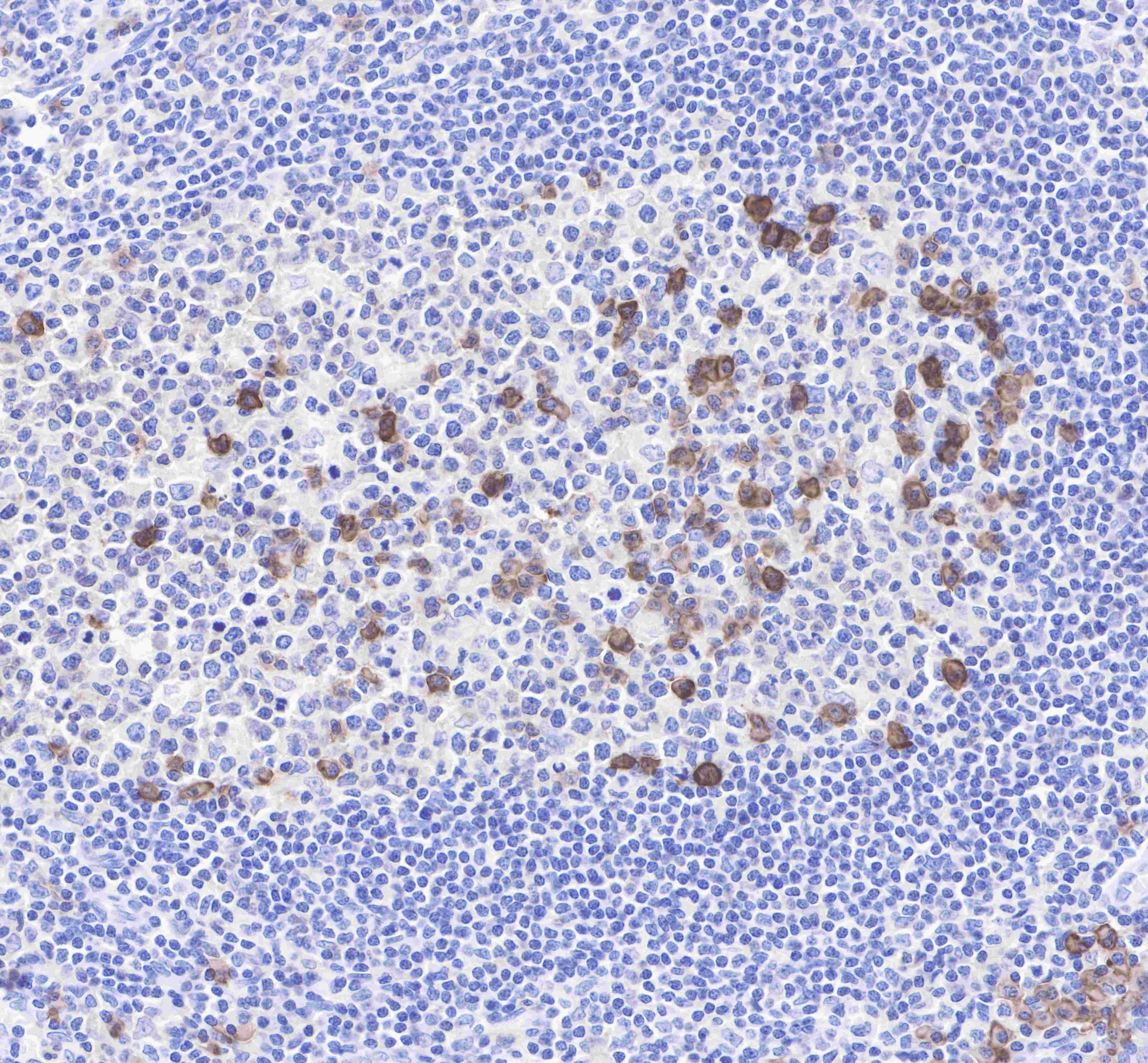
IHC shows positive staining in paraffin-embedded human tonsil. Anti-CD38 antibody was used at 1/1000 dilution, followed by a Goat Anti-Rabbit IgG H&L (HRP) ready to use. Counterstained with hematoxylin.
Heat mediated antigen retrieval with Tris/EDTA buffer pH9.0 was performed before commencing with IHC staining protocol.
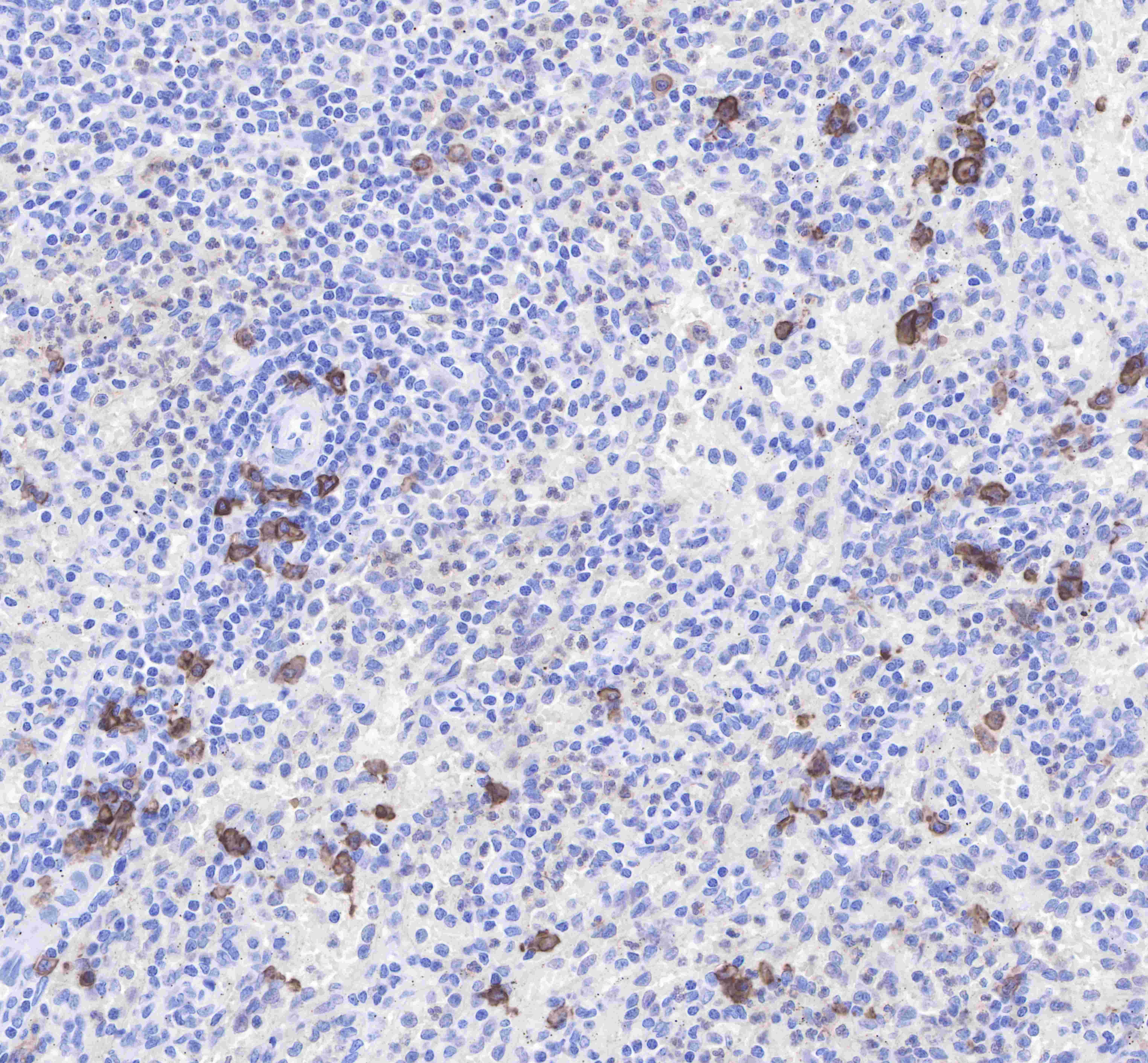
IHC shows positive staining in paraffin-embedded human spleen. Anti-CD38 antibody was used at 1/1000 dilution, followed by a Goat Anti-Rabbit IgG H&L (HRP) ready to use. Counterstained with hematoxylin.
Heat mediated antigen retrieval with Tris/EDTA buffer pH9.0 was performed before commencing with IHC staining protocol.
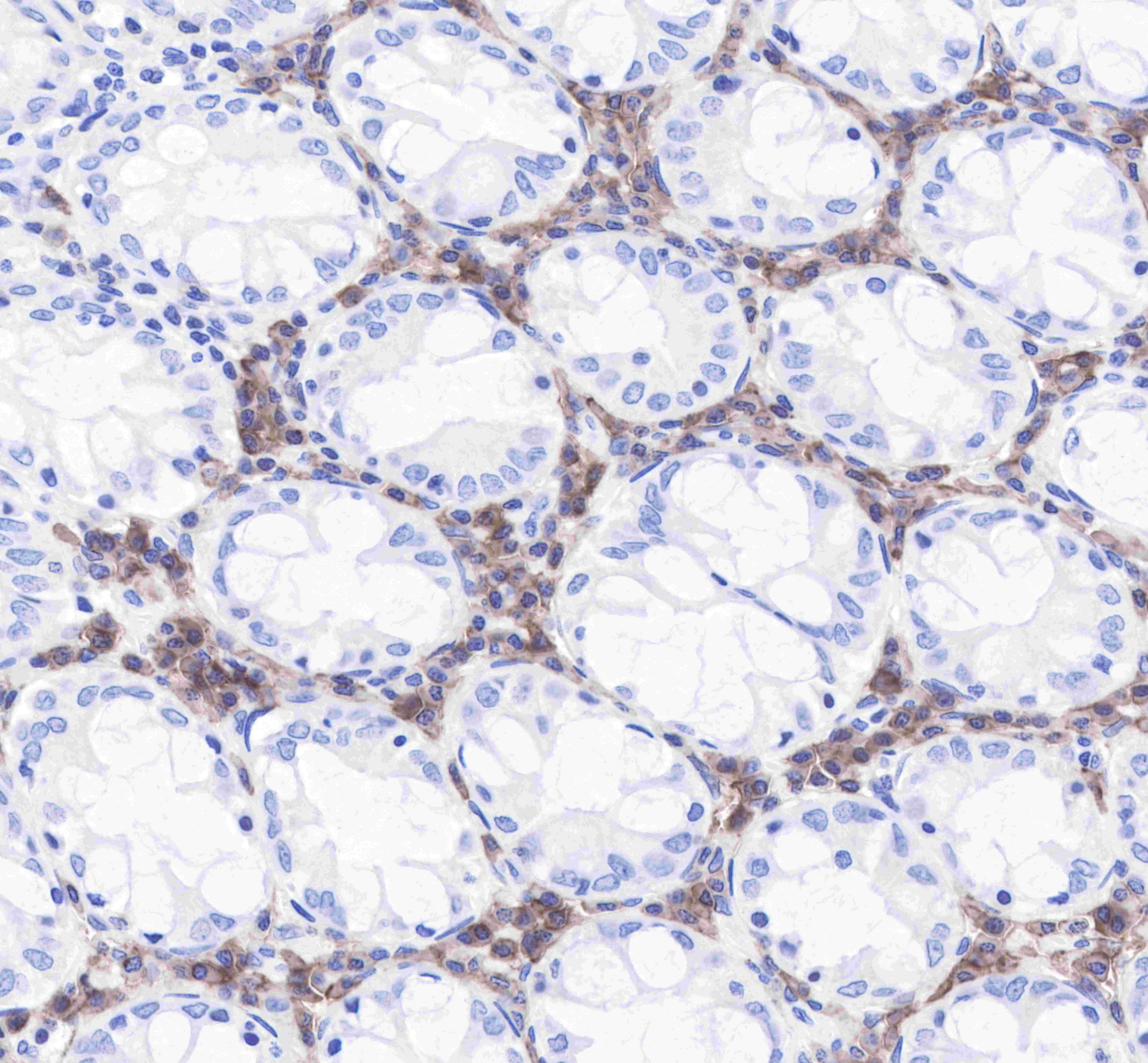
IHC shows positive staining in paraffin-embedded human colon. Anti-CD38 antibody was used at 1/1000 dilution, followed by a Goat Anti-Rabbit IgG H&L (HRP) ready to use. Counterstained with hematoxylin.
Heat mediated antigen retrieval with Tris/EDTA buffer pH9.0 was performed before commencing with IHC staining protocol.
IHC shows positive staining in paraffin-embedded human stomach. Anti-CD38 antibody was used at 1/1000 dilution, followed by a Goat Anti-Rabbit IgG H&L (HRP) ready to use. Counterstained with hematoxylin.
Heat mediated antigen retrieval with Tris/EDTA buffer pH9.0 was performed before commencing with IHC staining protocol.
IHC shows positive staining in paraffin-embedded human lung adenocarcinoma. Anti-CD38 antibody was used at 1/1000 dilution, followed by a Goat Anti-Rabbit IgG H&L (HRP) ready to use. Counterstained with hematoxylin.
Heat mediated antigen retrieval with Tris/EDTA buffer pH9.0 was performed before commencing with IHC staining protocol.
IHC shows positive staining in paraffin-embedded human prostate cancer. Anti-CD38 antibody was used at 1/1000 dilution, followed by a Goat Anti-Rabbit IgG H&L (HRP) ready to use. Counterstained with hematoxylin.
Heat mediated antigen retrieval with Tris/EDTA buffer pH9.0 was performed before commencing with IHC staining protocol.
免疫细胞化学
ICC shows positive staining in Ramos cells. Anti-CD38 antibody was used at 1/50 dilution (Green) and incubated overnight at 4°C. Goat polyclonal Antibody to Rabbit IgG - H&L (Alexa Fluor® 488) was used as secondary antibody at 1/1000 dilution. The cells were fixed with 4% PFA and permeabilized with 0.1% PBS-Triton X-100. Nuclei were counterstained with DAPI (Blue).
Negative control:ICC shows negative staining in HeLa cells. Anti-CD38 antibody was used at 1/50 dilution and incubated overnight at 4°C. Goat polyclonal Antibody to Rabbit IgG - H&L (Alexa Fluor® 488) was used as secondary antibody at 1/1000 dilution. The cells were fixed with 4% PFA and permeabilized with 0.1% PBS-Triton X-100. Nuclei were counterstained with DAPI (Blue).

评论(0)