


 申请试用
申请试用 产品介绍 评论(0)
宿主来源
Rabbit抗原名称
CD79B分子别名
B-cell-specific glycoprotein B29; Ig-beta; Immunoglobulin-associated B29 protein免疫原
Synthetic Peptide细胞定位
MembraneAccession
P40259克隆号
SDT-043-9抗体类型
Rabbit mAb应用
IHC-P ? ,ICC ? ,WB ,IP反应种属 ?
Hu, Ms纯化方式
Protein A浓度
0.5mg/ml分子量
30-50kDa
标记
Unconjugated性状
Liquid缓冲体系
PBS, 40% Glycerol, 0.05%BSA, 0.03% Proclin 300
储存条件
12 months from date of receipt / reconstitution, -20 °C as supplied
| 应用 | 稀释度 | 推荐种属 |
|---|---|---|
| IP | 1:25 | Hu |
| IHC-P | 1:2000 | Hu, Ms |
| ICC | 1:500 | Hu |
| WB | 1:1000 | Hu |
CD79B is a multimeric complex that includes the antigen-specific component, surface immunoglobulin (Ig). Surface Ig non-covalently associates with two other proteins, Ig-alpha and Ig-beta, which are necessary for expression and function of the B-cell antigen receptor. This gene encodes the Ig-beta protein of the B-cell antigen component. CD79B is required in cooperation with CD79A for initiation of the signal transduction cascade activated by the B-cell antigen receptor complex (BCR) which leads to internalization of the complex, trafficking to late endosomes and antigen presentation. And it also enhances phosphorylation of CD79A, possibly by recruiting kinases which phosphorylate CD79A or by recruiting proteins which bind to CD79A and protect it from dephosphorylation.
免疫印迹
WB result of CD79B Rabbit mAb
Primary antibody : CD79B Rabbit mAb at 1/1000 dilution
Lane 1: Jurkat whole cell lysate 20 µg
Lane 2: Raji whole cell lysate 20 µg
Lane 3: Daudi whole cell lysate 20 µg
Lane 4: Ramos whole cell lysate 20 µg
Negative control: Jurkat whole cell lysateSecondary antibody: Goat Anti-Rabbit IgG, (H+L), HRP conjugated at 1/10000 dilution
Predicted MW: 26 kDa
Observed MW: 35~40 kDa
Exposure time: 4s
免疫沉淀
CD79B Rabbit mAb at 1/25 dilution (2µg) immunoprecipitating CD79B in 0.4mg Daudi whole cell lysate. Western blot was performed on the immunoprecipitate using CD79B Rabbit mAb at 1/1000 dilution. Secondary antibody (HRP) for IP was used at 1/400 dilution.
Lane 1: Daudi whole cell lysate 10µg (input)
Lane 2: CD79B Rabbit mAb IP in Daudi whole cell lysate
Lane 3: Rabbit monoclonal IgG IP in Daudi whole cell lysate
Predicted MW: 26 kDa
Observed MW: 35~40 kDa
Exposure time: 8s
免疫组化
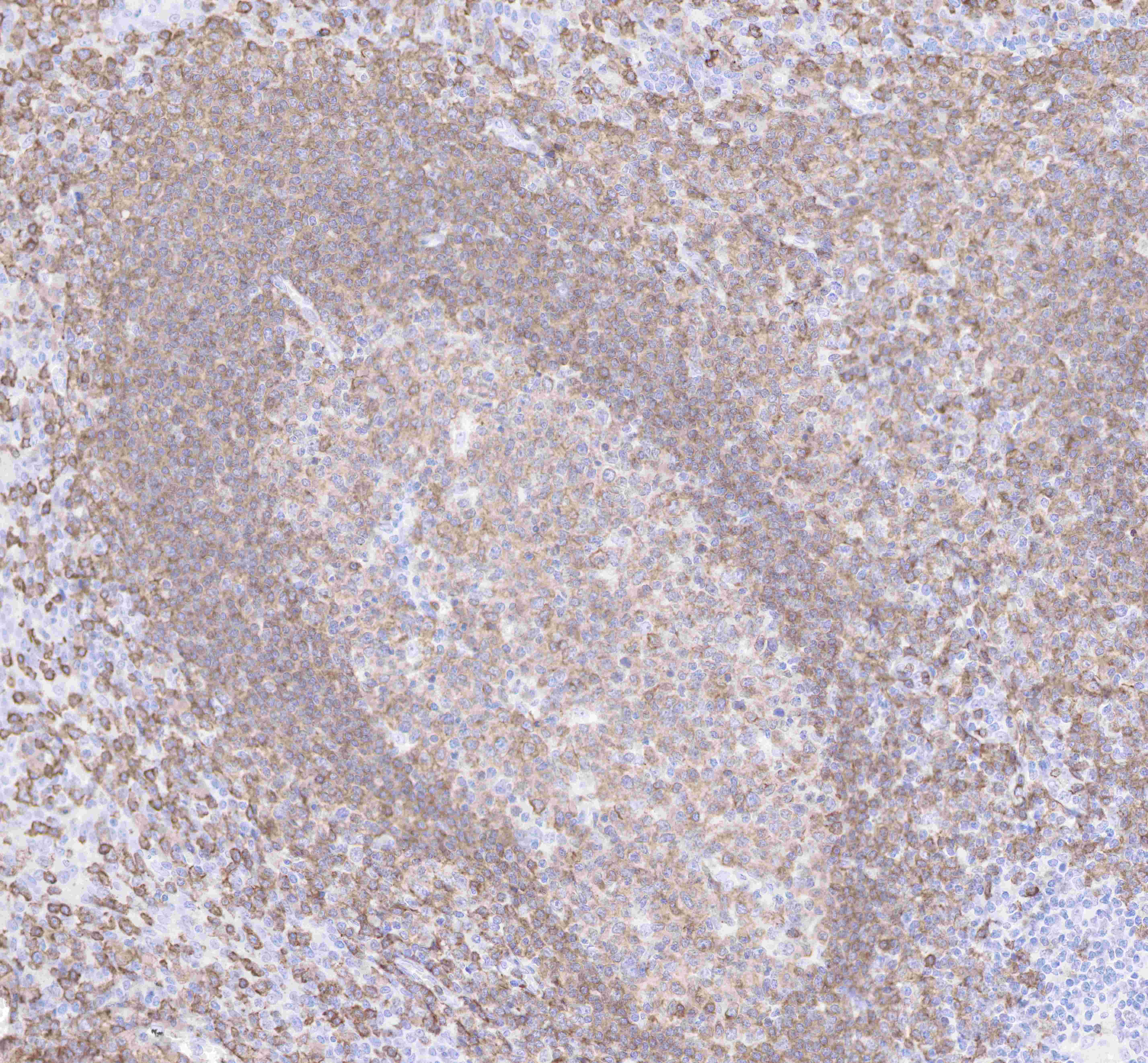
IHC shows positive staining in paraffin-embedded human tonsil.
Anti-CD79B antibody was used at 1/2000 dilution, followed by a Goat Anti-Rabbit IgG H&L (HRP) ready to use.
Counterstained with hematoxylin.
Heat mediated antigen retrieval with Tris/EDTA buffer pH9.0 was performed before commencing with IHC staining protocol.
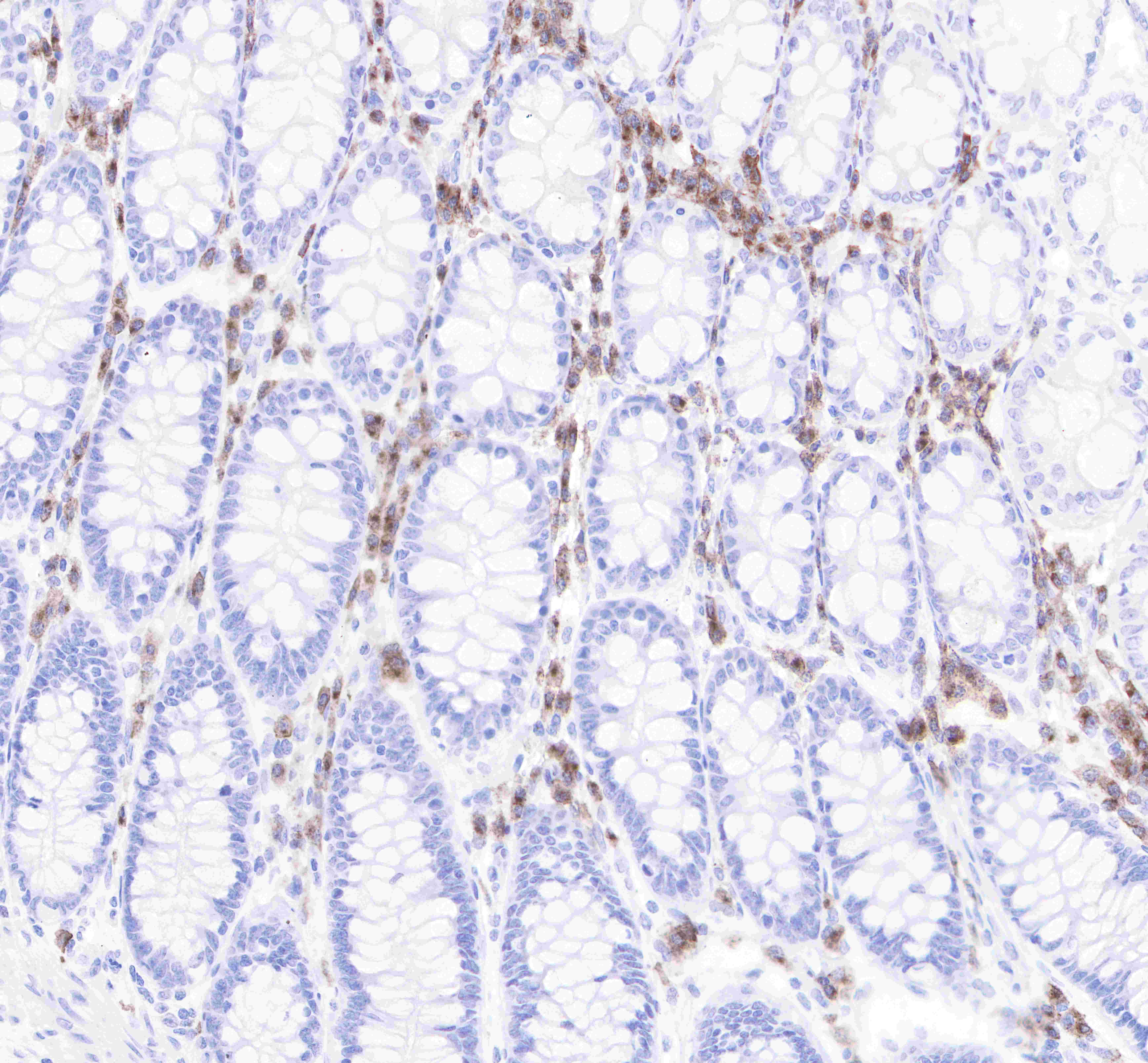
IHC shows positive staining in paraffin-embedded human colon.
Anti-CD79B antibody was used at 1/2000 dilution, followed by a Goat Anti-Rabbit IgG H&L (HRP) ready to use.
Counterstained with hematoxylin.
Heat mediated antigen retrieval with Tris/EDTA buffer pH9.0 was performed before commencing with IHC staining protocol.
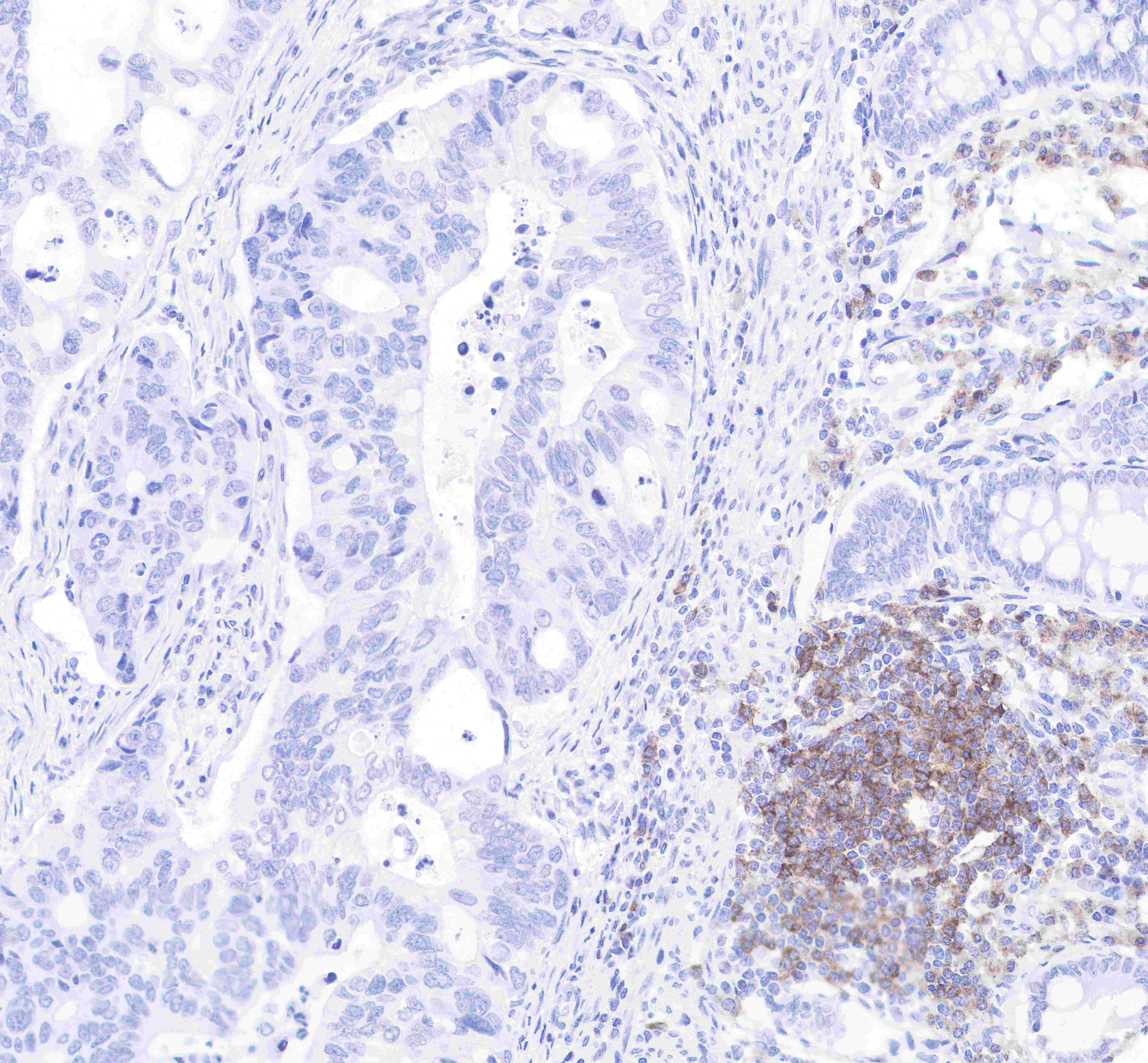
IHC shows positive staining in paraffin-embedded human colon cancer.
Anti-CD79B antibody was used at 1/2000 dilution, followed by a Goat Anti-Rabbit IgG H&L (HRP) ready to use.
Counterstained with hematoxylin.
Heat mediated antigen retrieval with Tris/EDTA buffer pH9.0 was performed before commencing with IHC staining protocol.
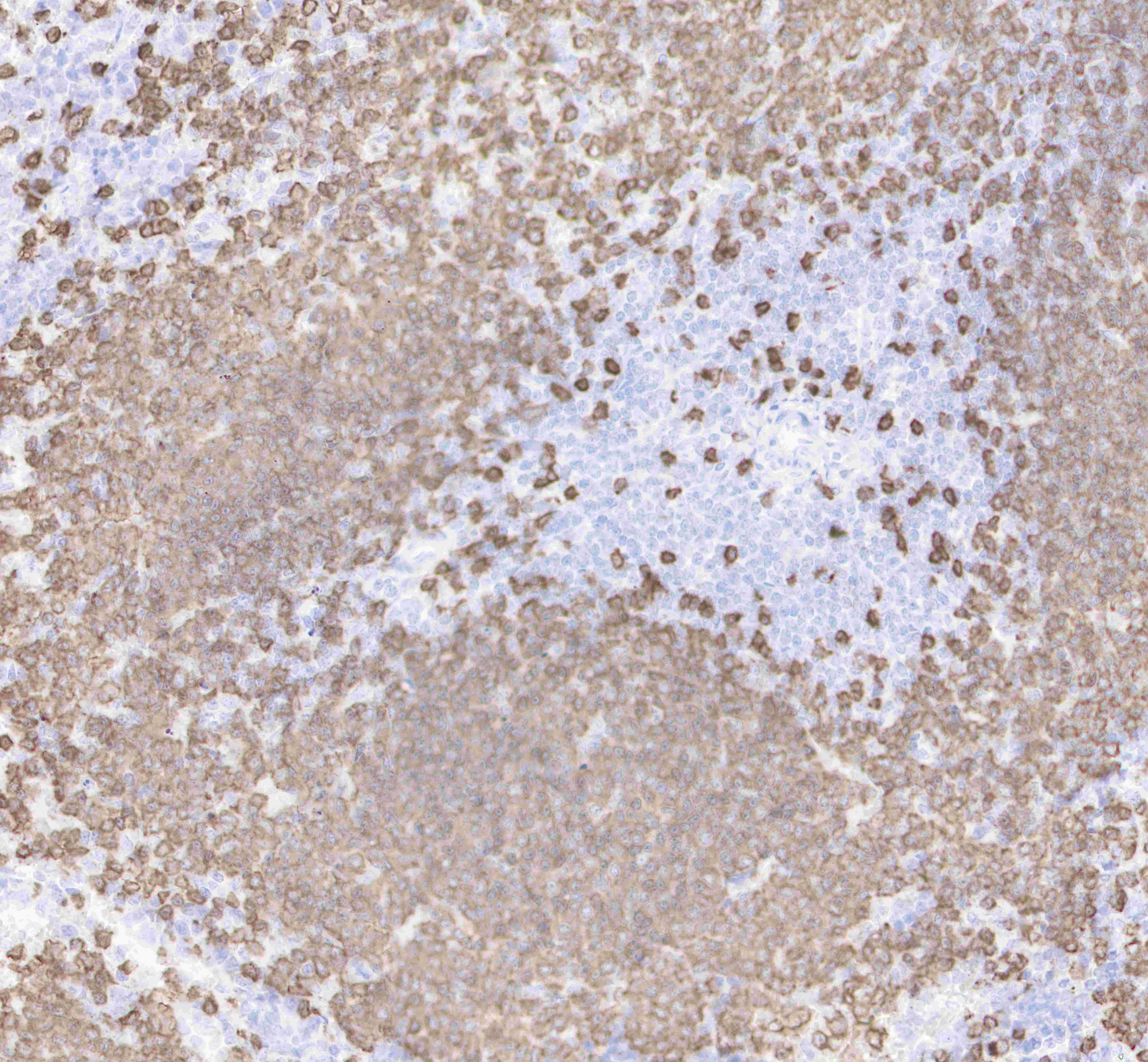
IHC shows positive staining in paraffin-embedded mosue spleen.
Anti-CD79B antibody was used at 1/2000 dilution, followed by a Goat Anti-Rabbit IgG H&L (HRP) ready to use.
Counterstained with hematoxylin.
Heat mediated antigen retrieval with Tris/EDTA buffer pH9.0 was performed before commencing with IHC staining protocol.
免疫细胞化学
ICC shows positive staining in Ramos cells (top panel) and negative staining in Jurkat cells (below panel). Anti-CD79B antibody was used at 1/500 dilution (Green) and incubated overnight at 4°C. Goat polyclonal Antibody to Rabbit IgG - H&L (Alexa Fluor® 488) was used as secondary antibody at 1/1000 dilution. The cells were fixed with 4% PFA and permeabilized with 0.1% PBS-Triton X-100. Nuclei were counterstained with DAPI (Blue). Counterstain with tubulin (Red).

评论(0)