




 分享
分享产品介绍 评论(0)
宿主来源
Rabbit抗原名称
Keratin 14分子别名
Cytokeratin-14,CK-14, KRT14免疫原
Synthetic PeptideAccession
P02533克隆号
SDT-023-1抗体类型
Rabbit mAb反应种属 ?
Hu, Ms纯化方式
Protein A研究领域
Signal Transduction浓度
1 mg/ml分子量
51kDa
标记
Unconjugated性状
Liquid缓冲体系
PBS
储存条件
12 months from date of receipt, 4°C as supplied
应用
IHC-P ?
ICC ?
WB
IP
稀释度
应用 稀释度 推荐种属 WB 1:1000 Hu, Ms IHC-P 1:1000 Hu, Ms ICC 1:500 Hu IP 1:25 Hu
Keratin 14 is a member of the type I keratin family of intermediate filament proteins. Keratin 14 was the first type I keratin sequence determined. Keratin 14 is also known as cytokeratin-14 (CK-14) or keratin-14 (KRT14). In humans it is encoded by the KRT14 gene. Keratin 14 is expressed in mitotically active basal layer cells, along with its partner keratin 5 (K5), and their expression is down-regulated as cells differentiate. Keratin 14 has been studied as a prognostic marker in breast cancer. Keratin 14 distinguishes stratified epithelial cells from simple epithelial cells and has been reported useful in the identification of squamous cell carcinomas.
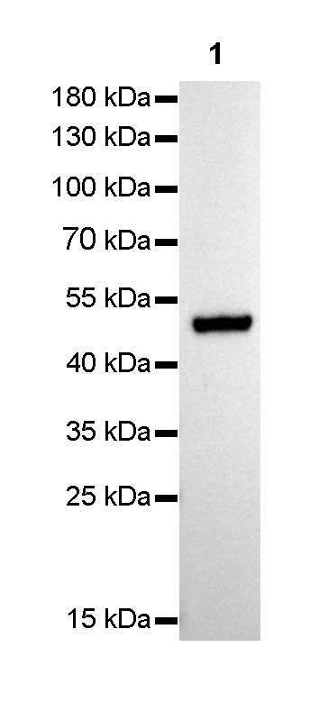
免疫印迹
WB result of Keratin 14 Rabbit mAb
Primary antibody : Keratin 14 Rabbit mAb at 1/1000 dilution
Lane 1 : A431 whole cell lysate 5 µg
Secondary antibody: Goat Anti-Rabbit IgG, (H+L), HRP conjugated at 1/10000 dilution
Predicted MW: 51 kDa
Observed MW: 51 kDa
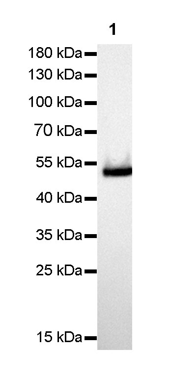
Exposure time: 0.9 secondWB result of Keratin 14 Rabbit mAb
Primary antibody : Keratin 14 Rabbit mAb at 1/1000 dilution
Lane 1 : mouse skin lysate 5 µg
Secondary antibody: Goat Anti-Rabbit IgG, (H+L), HRP conjugated at 1/10000 dilution
Predicted MW: 51 kDa
Observed MW: 53 kDa
Exposure time: 0.9 seconds
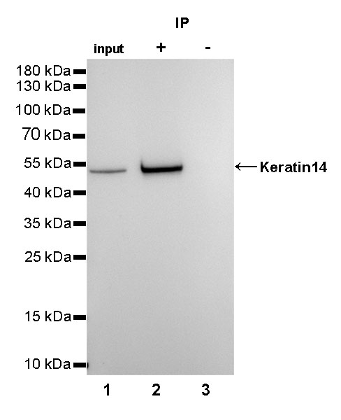
免疫沉淀
Keratin 14 Rabbit mAb at 1/25 dilution (2µg) immunoprecipitating Keratin 14 in 0.4mg A431 whole cell lysate. Western blot was performed on the immunoprecipitate using Keratin 14 Rabbit mAb at 1/1000 dilution. Secondary antibody (HRP) for IP was used at 1/400 dilution.
Lane 1: A431 whole cell lysate 10µg (input)
Lane 2: Keratin 14 Rabbit mAb IP in A431 whole cell lysate
Lane 3: Rabbit monoclonal IgG IP in A431 whole cell lysate
Predicted MW: 51 kDa
Observed MW: 51 kDa
Exposure time: 3s
免疫组化
IHC shows membrane staining in paraffin-embedded human skin squamous epithelium.
Anti-Keratin 14 antibody was used at 1/1000 dilution, followed by a Goat Anti-Rabbit IgG H&L (HRP) ready to use.
Counterstained with hematoxylin.
Heat mediated antigen retrieval with Tris/EDTA buffer pH9.0 was performed before commencing with IHC staining protocol.
IHC shows membrane staining in paraffin-embedded human tonsil squamous epithelium.
Anti-Keratin 14 antibody was used at 1/1000 dilution, followed by a Goat Anti-Rabbit IgG H&L (HRP) ready to use.
Counterstained with hematoxylin.
Heat mediated antigen retrieval with Tris/EDTA buffer pH9.0 was performed before commencing with IHC staining protocol.
IHC shows membrane staining in paraffin-embedded human prostate.
Anti-Keratin 14 antibody was used at 1/1000 dilution, followed by a Goat Anti-Rabbit IgG H&L (HRP) ready to use.
Counterstained with hematoxylin.
Heat mediated antigen retrieval with Tris/EDTA buffer pH9.0 was performed before commencing with IHC staining protocol.
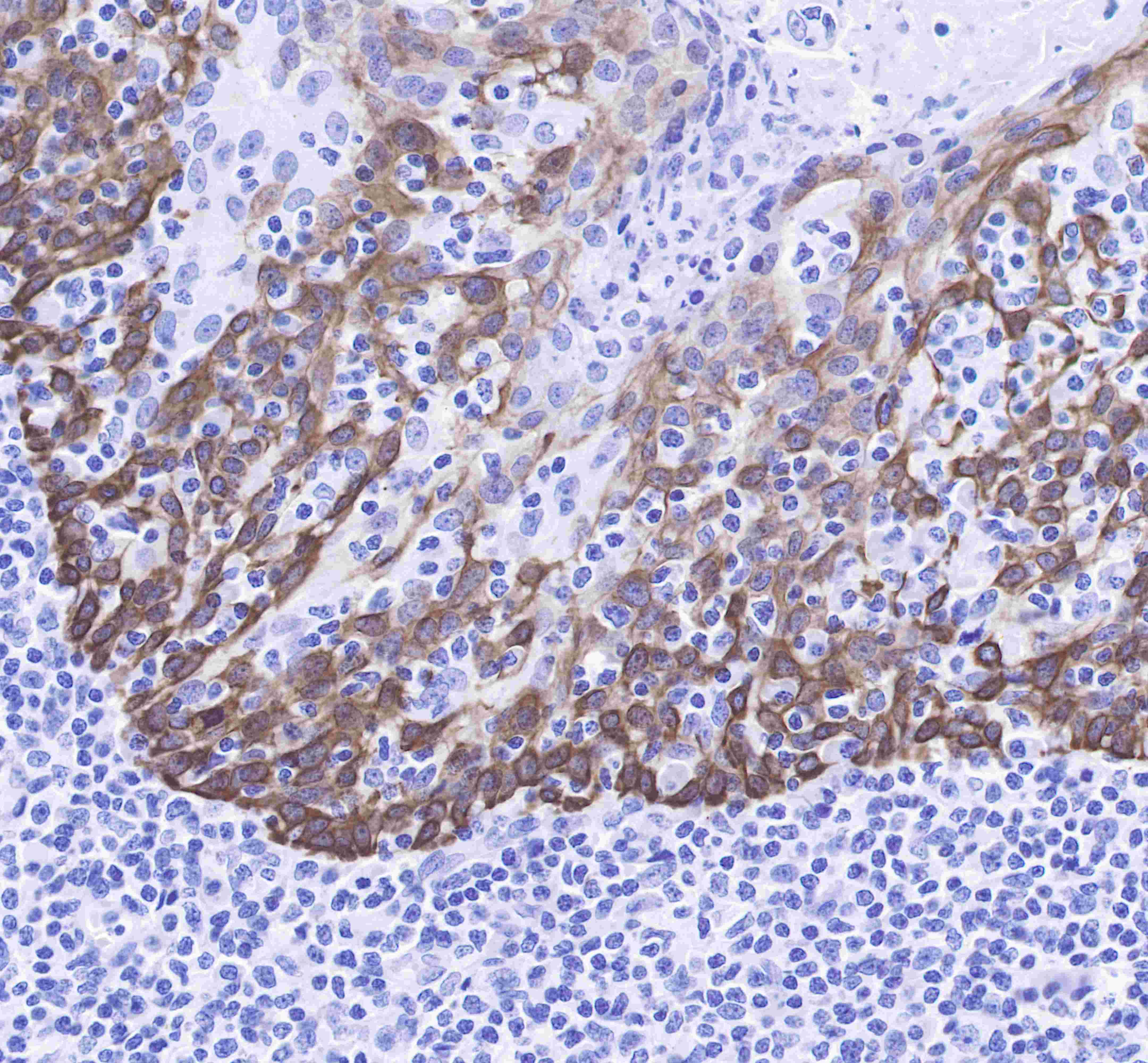
IHC shows membrane staining in paraffin-embedded human cervix cancer. Anti-Keratin 14 antibody was used at 1/1000 dilution, followed by a Goat Anti-Rabbit IgG H&L (HRP) ready to use.
Counterstained with hematoxylin.
Heat mediated antigen retrieval with Tris/EDTA buffer pH9.0 was performed before commencing with IHC staining protocol.
IHC shows membrane staining in paraffin-embedded human lung squamous cell cancer.
Anti-Keratin 14 antibody was used at 1/1000 dilution, followed by a Goat Anti-Rabbit IgG H&L (HRP) ready to use.
Counterstained with hematoxylin.
Heat mediated antigen retrieval with Tris/EDTA buffer pH9.0 was performed before commencing with IHC staining protocol.
IHC shows membrane staining in paraffin-embedded mouse skin squamous epithelium.
Anti-Keratin 14 antibody was used at 1/1000 dilution, followed by a Goat Anti-Rabbit IgG H&L (HRP) ready to use.
Counterstained with hematoxylin.
Heat mediated antigen retrieval with Tris/EDTA buffer pH9.0 was performed before commencing with IHC staining protocol.
免疫细胞化学
ICC shows membrane staining in A431 cells.
Anti-Keratin 14 antibody was used at 1/500 dilution and incubated overnight at 4°C.
Goat polyclonal Antibody to Rabbit IgG - H&L (Alexa Fluor® 488) was used as secondary antibody at 1/1000 dilution.
The cells were fixed with 100% methanol and permeabilized with 0.1% PBS-Triton X-100.
Nuclei were counterstained with DAPI.

评论(0)