




 分享
分享产品介绍 评论(0)
宿主来源
Rabbit抗原名称
CD31分子别名
PECAM-1,EndoCAM, GPIIA, PECA1免疫原
Synthetic PeptideAccession
P16284克隆号
SDT-008-17抗体类型
Rabbit mAb抗体同种型
IgG反应种属 ?
Hu纯化方式
Protein A研究领域
Immunology浓度
0.5mg/ml分子量
150kDa
标记
Unconjugated性状
Liquid缓冲体系
PBS, 40% Glycerol, 0.05%BSA, 0.03% Proclin 300
储存条件
12 months from date of receipt / reconstitution, -20 °C as supplied
应用
IHC-P ?
WB
IP
稀释度
应用 稀释度 IHC-P 1:250 IP 1:25 WB 1:1000
Cluster of differentiation 31 (CD31) also known as platelet endothelial cell adhesion molecule (PECAM-1) is a member of the immunoglobulin superfamily and is likely involved in leukocyte transmigration, angiogenesis, and integrin activation. PECAM-1 plays a key role in removing aged neutrophils from the body. PECAM-1 is found on the surface of platelets, monocytes, neutrophils, and some types of T-cells, and makes up a large portion of endothelial cell intercellular junctions. In immunohistochemistry, CD31 is used primarily to demonstrate the presence of endothelial cells in histological tissue sections. This can help to evaluate the degree of tumor angiogenesis, which can imply a rapidly growing tumor. Malignant endothelial cells also commonly retain the antigen, so that CD31 immunohistochemistry can also be used to demonstrate both angiomas and angiosarcomas. It can also be demonstrated in small lymphocytic and lymphoblastic lymphomas, although more specific markers are available for these conditions.
免疫印迹
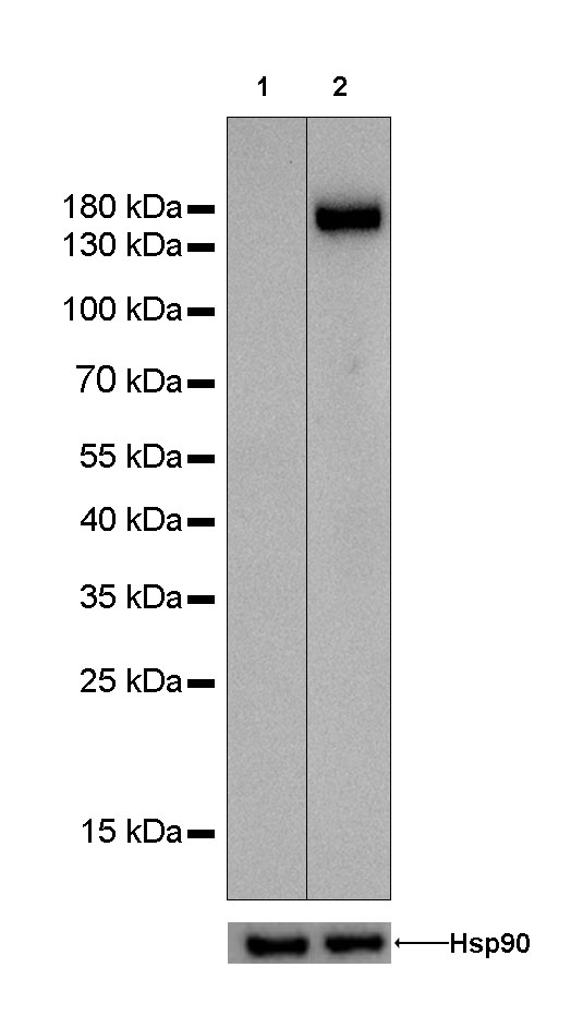
WB result of anti-CD31 antibody
Primary antibody : Anti-CD31 antibody at 1/1000 dilution
Lane 1 : Hela whole cell lysate 20 µg
Lane 2 : THP1 whole cell lysate 20 µg
Negative control: Hela whole cell lysateSecondary antibody: Goat Anti-Rabbit IgG, (H+L), HRP conjugated at 1/10000 dilution
Predicted MW: 82 kDa
Observed MW: 150 kDa
免疫沉淀
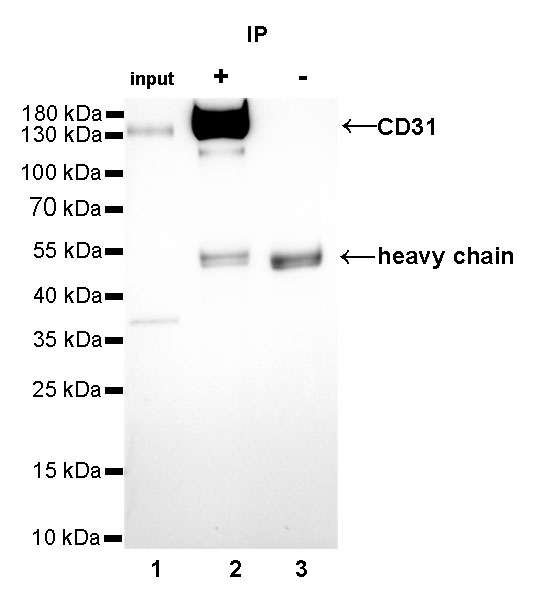
CD31 Rabbit mAb at 1/25 dilution (2µg) immunoprecipitating CD31 in 0.4mg THP-1 whole cell lysate. Western blot was performed on the immunoprecipitate using CD31 Rabbit mAb at 1/1000 dilution. Secondary antibody (HRP) for IP was used at 1/400 dilution.
Lane 1: THP-1 whole cell lysate 10µg (input)
Lane 2: CD31 Rabbit mAb IP in THP-1 whole cell lysate
Lane 3: Rabbit monoclonal IgG IP in THP-1 whole cell lysate
Predicted MW: 82 kDa
Observed MW: 150 kDa
免疫组化
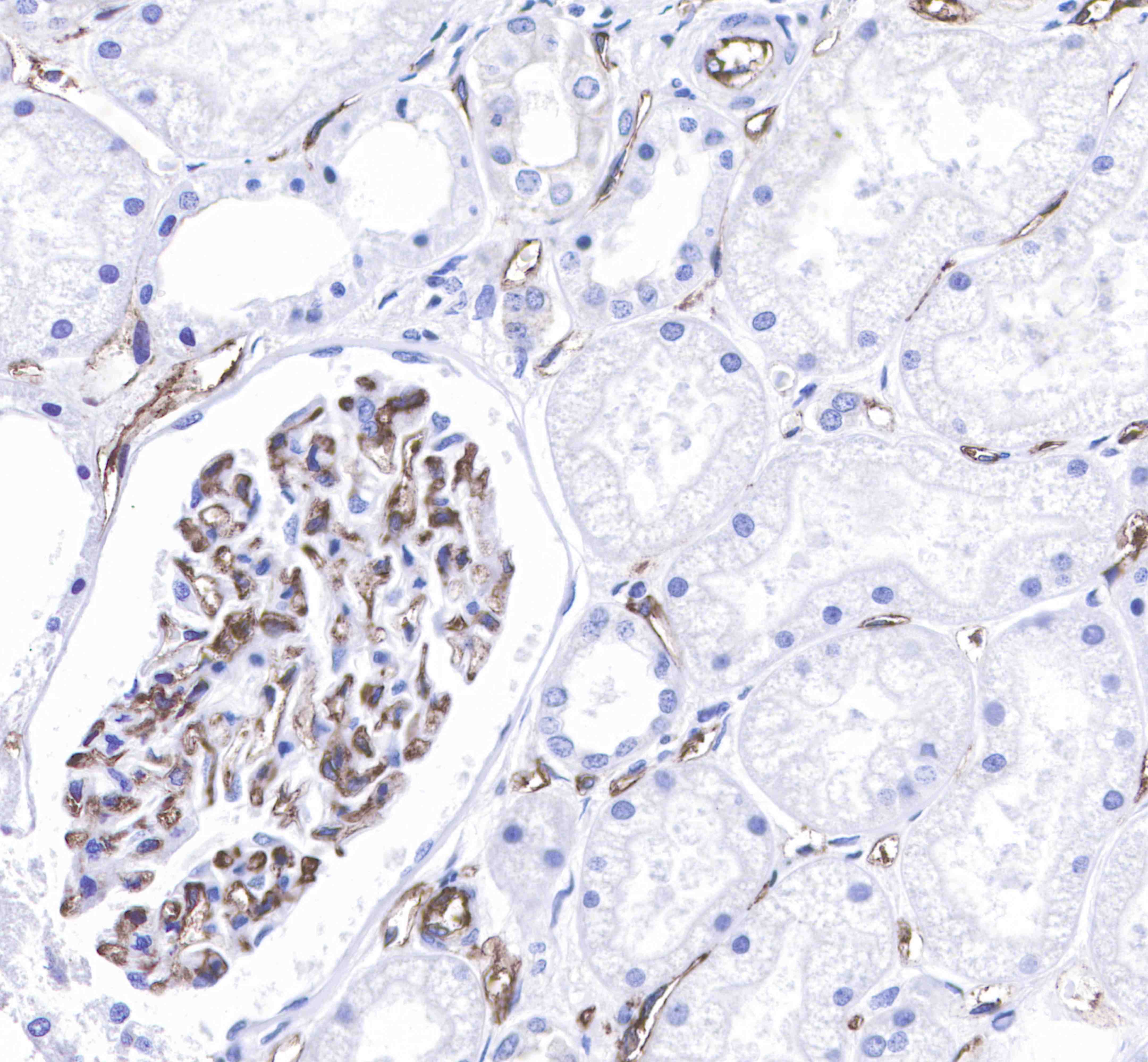
IHC shows membrane staining in paraffin-embedded human kidney endothelial cells.
Anti-CD31 antibody was used at 1/250 dilution, followed by a Goat Anti-Rabbit IgG H&L (HRP) ready to use.
Counterstained with hematoxylin.
Heat mediated antigen retrieval with Tris/EDTA buffer pH9.0 was performed before commencing with IHC staining protocol.
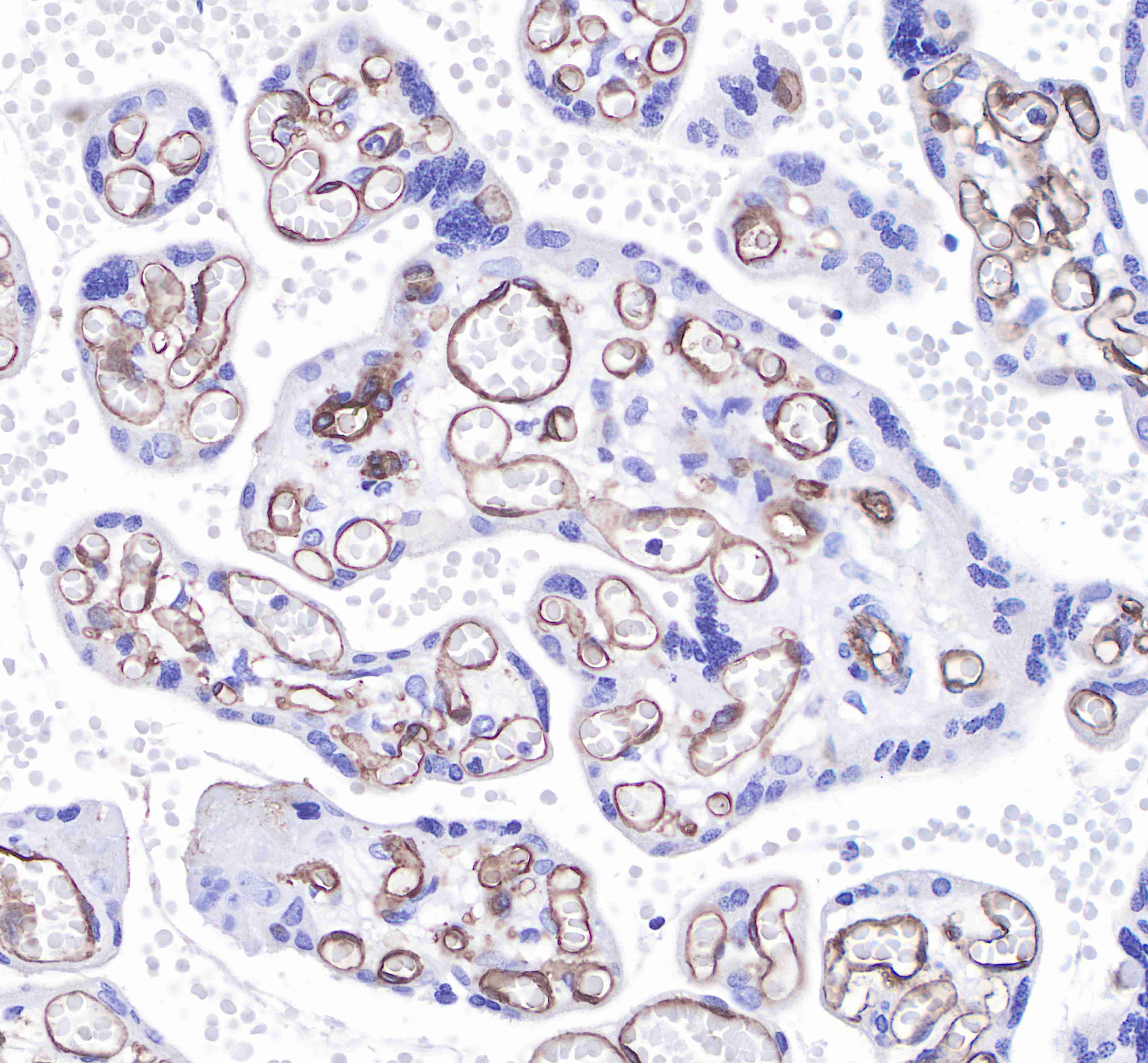
IHC shows membrane staining in paraffin-embedded human placenta endothelial cells.
Anti-CD31 antibody was used at 1/250 dilution, followed by a Goat Anti-Rabbit IgG H&L (HRP) ready to use.
Counterstained with hematoxylin.
Heat mediated antigen retrieval with Tris/EDTA buffer pH9.0 was performed before commencing with IHC staining protocol.
IHC shows membrane staining in paraffin-embedded human lung squamous cell cancer endothelial cells.
Anti-CD31 antibody was used at 1/250 dilution, followed by a Goat Anti-Rabbit IgG H&L (HRP) ready to use.
Counterstained with hematoxylin.
Heat mediated antigen retrieval with Tris/EDTA buffer pH9.0 was performed before commencing with IHC staining protocol.
IHC shows membrane staining in paraffin-embedded human hepatocellular carcinoma endothelial cells.
Anti-CD31 antibody was used at 1/250 dilution, followed by a Goat Anti-Rabbit IgG H&L (HRP) ready to use.
Counterstained with hematoxylin.
Heat mediated antigen retrieval with Tris/EDTA buffer pH9.0 was performed before commencing with IHC staining protocol.

评论(0)