


 大包装询价
大包装询价 产品介绍 评论(0)
宿主来源
Rabbit抗原名称
HexokinaseⅠ分子别名
Brain form hexokinase; Hexokinase type I (HK I); Hexokinase-A; HK1免疫原
Synthetic Peptide细胞定位
Mitochondrion outer membraneAccession
P19367克隆号
S-868-113抗体类型
Recombinant mAb抗体同种型
IgG应用
ICFCM, ICC, WB反应种属 ?
Hu, Ms阳性样本
293T, HeLa, Jurkat, MCF7, U-87 MG, NIH/3T3, C2C12纯化方式
Protein A浓度
0.5 mg/ml标记
Unconjugated性状
Liquid缓冲体系
PBS, 40% Glycerol, 0.05% BSA, 0.03% Proclin 300
储存条件
12 months from date of receipt / reconstitution, -20 °C as supplied
| 应用 | 稀释度 | 推荐种属 |
|---|---|---|
| WB | 1:1000 | Hu, Ms |
| ICC | 1:500 | Hu |
| ICFCM | 1:50 |
Hexokinase I (HK-I) is a subtype of the hexokinase family. The primary function of HK-I is to regulate the entry of glucose into the glycolytic pathway and generate ATP. It is firmly bound to mitochondria and, in the brain, HK-I catalyzes the phosphorylation of glucose, determining the rate of glycolysis in the brain. HK-I also acts as a coordinating enzyme to prevent an imbalance between glycolysis and mitochondrial oxidative reactions, which could lead to the accumulation of lactic acid. Additionally, in hematopoietic cell lines, HK-I is co-expressed with GLUT1, increasing the level of glucose phosphorylation and the level of cellular nicotinamide adenine dinucleotide phosphate (NADPH), thereby exerting an anti-apoptotic effect.
免疫印迹
WB result of HexokinaseⅠRecombinant Rabbit mAb
Primary antibody: HexokinaseⅠRecombinant Rabbit mAb at 1/1000 dilution
Lane 1: 293T whole cell lysate 20 µg
Lane 2: HeLa whole cell lysate 20 µg
Lane 3: Jurkat whole cell lysate 20 µg
Lane 4: MCF7 whole cell lysate 20 µg
Lane 5: U-87 MG whole cell lysate 20 µg
Secondary antibody: Goat Anti-Rabbit IgG, (H+L), HRP conjugated at 1/10000 dilution Predicted MW: 102 kDa
Observed MW: 110 kDaWB result of HexokinaseⅠRecombinant Rabbit mAb
Primary antibody: HexokinaseⅠRecombinant Rabbit mAb at 1/1000 dilution
Lane 1: NIH/3T3 whole cell lysate 20 µg
Lane 2: C2C12 whole cell lysate 20 µg
Secondary antibody: Goat Anti-Rabbit IgG, (H+L), HRP conjugated at 1/10000 dilution Predicted MW: 102 kDa
Observed MW: 110 kDa
流式分析
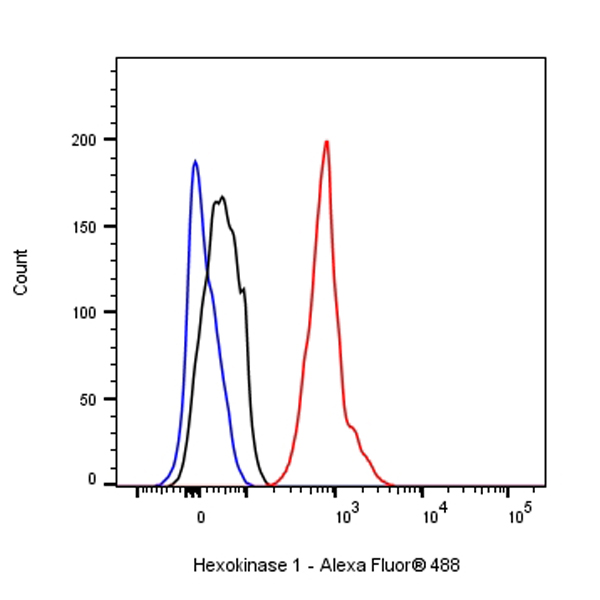
Flow cytometric analysis of 4% PFA fixed 90% methanol permeabilized HeLa (Human cervix adenocarcinoma epithelial cell) cells labelling Hexokinase 1 antibody at 1/50 dilution (1 μg)/ (Red) compared with a Rabbit monoclonal IgG (Black) isotype control and an unlabelled control (cells without incubation with primary antibody and secondary antibody) (Blue). Goat Anti - Rabbit IgG Alexa Fluor® 488 was used as the secondary antibody.
免疫细胞化学
ICC shows positive staining in HeLa cells. Anti- Hexokinase 1 antibody was used at 1/500 dilution (Green) and incubated overnight at 4°C. Goat polyclonal Antibody to Rabbit IgG - H&L (Alexa Fluor® 488) was used as secondary antibody at 1/1000 dilution. The cells were fixed with 100% ice-cold methanol and permeabilized with 0.1% PBS-Triton X-100. Nuclei were counterstained with DAPI (Blue). Counterstain with tubulin (Red).

评论(0)