


p38α MAPK+p38β MAPK Recombinant Rabbit mAb (S-1300-102)
Mitogen-activated protein kinase 14,MAP kinase 14,MAPK 14,Cytokine suppressive anti-inflammatory drug-binding protein (CSAID-binding protein,CSBP),MAP kinase MXI2,MAX-interacting protein 2,Mitogen-activated protein kinase p38 alpha (MAP kinase p38 alpha),Stress-activated protein kinase 2a (SAPK2a),CSBP,CSBP1,CSBP2,CSPB1,MXI2,SAPK2A,Mitogen-activated protein kinase 11,MAP kinase 11,MAPK 11,Mitogen-activated protein kinase p38 beta (MAP kinase p38 beta,p38b),Stress-activated protein kinase 2b (SAPK2b),p38-2,PRKM11,SAPK2,SAPK2B
货号: S0B0895
- 价格: ¥260
- 规格:
- 数量:
 大包装询价
大包装询价 产品介绍 评论(0)
宿主来源
Rabbit抗原名称
p38α MAPK+p38β MAPK分子别名
Mitogen-activated protein kinase 14; MAP kinase 14; MAPK 14; Cytokine suppressive anti-inflammatory drug-binding protein (CSAID-binding protein; CSBP); MAP kinase MXI2; MAX-interacting protein 2; Mitogen-activated protein kinase p38 alpha (MAP kinase p38 alpha); Stress-activated protein kinase 2a (SAPK2a); CSBP; CSBP1; CSBP2; CSPB1; MXI2; SAPK2A; Mitogen-activated protein kinase 11; MAP kinase 11; MAPK 11; Mitogen-activated protein kinase p38 beta (MAP kinase p38 beta; p38b); Stress-activated protein kinase 2b (SAPK2b); p38-2; PRKM11; SAPK2; SAPK2B免疫原
Synthetic PeptideAccession
Q16539, Q15759克隆号
S-1300-102抗体类型
Recombinant mAb抗体同种型
IgG应用
ICFCM, IHC-P, ICC, WB, IP反应种属 ?
Hu, Ms, Rt预测反应种属
(反应种属缩写表)Cz, Zf, Bv纯化方式
Protein A浓度
0.5 mg/ml标记
Unconjugated性状
Liquid缓冲体系
PBS, 40% Glycerol, 0.05% BSA, 0.03% Proclin 300
储存条件
12 months from date of receipt / reconstitution, -20 °C as supplied
| 应用 | 稀释度 |
|---|---|
| WB | 1:1000 |
| IP | 1:50 |
| IHC-P | 1:200-1:1000 |
| ICC | 1:500 |
| ICFCM | 1:500 |
p38 MAPK, a member of the mitogen-activated protein kinase (MAPK) family, is activated by a variety of environmental stresses and inflammatory cytokines. p38 MAPK family includes p38α, p38β, p38γ, and p38δ. This pathway plays a crucial role in the regulation of cellular responses to stress, inflammation, and immune function. The p38 MAPK pathway is initiated by MAPK kinase kinases (MAPKKKs), such as MEKK or MLK, which phosphorylate and activate MAPK kinases (MAPKKs) like MKK3/6. These, in turn, phosphorylate and activate p38 MAPK. Downstream of p38 MAPK, a number of substrates are regulated, including HSP27, MAPKAPK-2 (MK2), MAPKAPK-3 (MK3), and various transcription factors like ATF-2, Stat1, Max/Myc complex, MEF-2, and Elk-1. This signaling pathway is involved in a wide range of biological processes, from the regulation of gene expression to cell cycle control, apoptosis, and autophagy. The p38 MAPK pathway is also implicated in the pathogenesis of various diseases, including cancer, where it can act as either a promoter or suppressor of tumor growth and metastasis, depending on the cellular context.
免疫印迹
WB result of p38α MAPK+p38β MAPK Recombinant Rabbit mAb
Primary antibody: p38α MAPK+p38β MAPK Recombinant Rabbit mAb at 1/1000 dilution
Lane 1: HeLa whole cell lysate 20 µg
Lane 2: Jurkat whole cell lysate 20 µg
Lane 3: MCF7 whole cell lysate 20 µg
Secondary antibody: Goat Anti-rabbit IgG, (H+L), HRP conjugated at 1/10000 dilution
Predicted MW: 41 kDa
Observed MW: 38 kDaWB result of p38α MAPK+p38β MAPK Recombinant Rabbit mAb
Primary antibody: p38α MAPK+p38β MAPK Recombinant Rabbit mAb at 1/1000 dilution
Lane 1: NIH/3T3 whole cell lysate 20 µg
Lane 2: mouse spleen lysate 20 µg
Secondary antibody: Goat Anti-rabbit IgG, (H+L), HRP conjugated at 1/10000 dilution
Predicted MW: 41 kDa
Observed MW: 38 kDaWB result of p38α MAPK+p38β MAPK Recombinant Rabbit mAb
Primary antibody: p38α MAPK+p38β MAPK Recombinant Rabbit mAb at 1/1000 dilution
Lane 1: C6 whole cell lysate 20 µg
Lane 2: rat spleen lysate 20 µg
Secondary antibody: Goat Anti-rabbit IgG, (H+L), HRP conjugated at 1/10000 dilution
Predicted MW: 41 kDa
Observed MW: 38 kDa
流式分析
Flow cytometric analysis of 4% PFA fixed 90% methanol permeabilized HeLa (Human cervix adenocarcinoma epithelial cell) labelling p38α MAPK+p38β MAPK antibody at 1/500 dilution (0.1 μg)/ (Red) compared with a Rabbit monoclonal IgG (Black) isotype control and an unlabelled control (cells without incubation with primary antibody and secondary antibody) (Blue). Goat Anti - Rabbit IgG Alexa Fluor® 488 was used as the secondary antibody.
Flow cytometric analysis of 4% PFA fixed 90% methanol permeabilized NIH/3T3 (Mouse embryonic fibroblast) labelling p38α MAPK+p38β MAPK antibody at 1/500 dilution (0.1 μg)/ (Red) compared with a Rabbit monoclonal IgG (Black) isotype control and an unlabelled control (cells without incubation with primary antibody and secondary antibody) (Blue). Goat Anti - Rabbit IgG Alexa Fluor® 488 was used as the secondary antibody.
免疫沉淀
p38α MAPK+p38β MAPK Rabbit mAb at 1/50 dilution (1 µg) immunoprecipitating p38α MAPK+p38β MAPK in 0.4 mg HeLa whole cell lysate.
Western blot was performed on the immunoprecipitate using p38α MAPK+p38β MAPK Rabbit mAb at 1/1000 dilution.
Secondary antibody (HRP) for IP was used at 1/1000 dilution.
Lane 1: HeLa whole cell lysate 20 µg (Input)
Lane 2: p38α MAPK+p38β MAPK Rabbit mAb IP in HeLa whole cell lysate
Lane 3: Rabbit monoclonal IgG IP in HeLa whole cell lysate
Predicted MW: 41 kDa
Observed MW: 38 kDa
免疫组化
IHC shows positive staining in paraffin-embedded human kidney. Anti-p38α MAPK+p38β antibody was used at 1/1000 dilution, followed by a HRP Polymer for Mouse & Rabbit IgG (ready to use). Counterstained with hematoxylin. Heat mediated antigen retrieval with Tris/EDTA buffer pH9.0 was performed before commencing with IHC staining protocol.
IHC shows positive staining in paraffin-embedded human colon cancer. Anti-p38α MAPK+p38β antibody was used at 1/200 dilution, followed by a HRP Polymer for Mouse & Rabbit IgG (ready to use). Counterstained with hematoxylin. Heat mediated antigen retrieval with Tris/EDTA buffer pH9.0 was performed before commencing with IHC staining protocol.
免疫细胞化学
ICC shows positive staining in HeLa cells. Anti-p38α MAPK+p38β MAPK antibody was used at 1/500 dilution (Green) and incubated overnight at 4°C. Goat polyclonal Antibody to Rabbit IgG - H&L (Alexa Fluor® 488) was used as secondary antibody at 1/1000 dilution. The cells were fixed with 4% PFA and permeabilized with 0.1% PBS-Triton X-100. Nuclei were counterstained with DAPI (Blue). Counterstain with tubulin (Red).
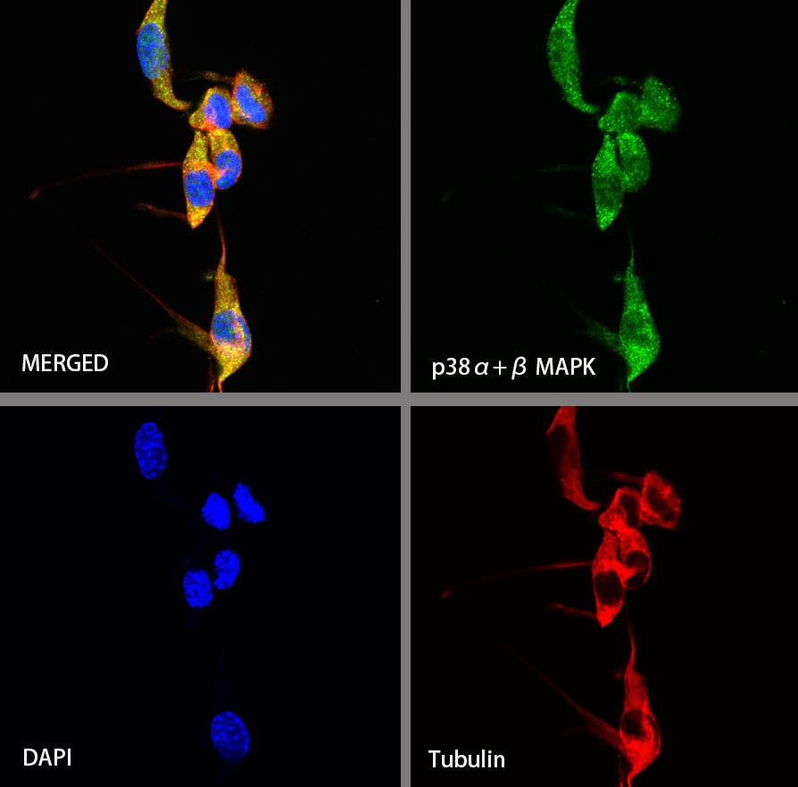
ICC shows positive staining in NIH/3T3 cells. Anti-p38α MAPK+p38β MAPK antibody was used at 1/500 dilution (Green) and incubated overnight at 4°C. Goat polyclonal Antibody to Rabbit IgG - H&L (Alexa Fluor® 488) was used as secondary antibody at 1/1000 dilution. The cells were fixed with 4% PFA and permeabilized with 0.1% PBS-Triton X-100. Nuclei were counterstained with DAPI (Blue). Counterstain with tubulin (Red).

评论(0)