


Galectin-1 Recombinant Rabbit mAb (S-1221-32)
LEG1,Galectin-1,Gal-1,14 kDa laminin-binding protein,HLBP14,14 kDa lectin,Beta-galactoside-binding lectin L-14-I,Galaptin,HBL,HPL,Lactose-binding lectin 1,Lectin galactoside-binding soluble 1,Putative MAPK-activating protein PM12,S-Lac lectin 1,LGALS1,Galectin-1/LGALS1,14 kDa laminin-binding protein (HLBP14)
货号: S0B0828
- 价格: ¥600
- 规格:
- 数量:
 大包装询价
大包装询价 产品介绍 评论(0)
别名
LEG1,Galectin-1,Gal-1,14 kDa laminin-binding protein,HLBP14,14 kDa lectin,Beta-galactoside-binding lectin L-14-I,Galaptin,HBL,HPL,Lactose-binding lectin 1,Lectin galactoside-binding soluble 1,Putative MAPK-activating protein PM12,S-Lac lectin 1,LGALS1,Galectin-1/LGALS1,14 kDa laminin-binding protein (HLBP14)宿主来源
Rabbit抗原名称
Galectin-1免疫原
Synthetic Peptide细胞定位
Cytoplasm, SecretedAccession
P09382克隆号
S-1221-32抗体类型
Recombinant mAb抗体同种型
IgG应用
IHC-P, ICC, WB, IP反应种属 ?
Hu, Ms, Rt纯化方式
Protein A浓度
0.5 mg/ml标记
Unconjugated性状
Liquid缓冲体系
PBS, 40% Glycerol, 0.05% BSA, 0.03% Proclin 300储存条件
12 months from date of receipt / reconstitution, -20 °C as supplied
| 应用 | 稀释度 |
|---|---|
| WB | 1:1000 |
| IHC-P | 1:1000 |
| ICC | 1:500 |
| IP | 1:50 |
Galectin-1, a member of the galectin family of proteins, is a versatile and multifunctional molecule that plays pivotal roles in various biological processes. Galectin-1 supports tumor growth and metastasis by promoting tumor angiogenesis, inhibiting T-cell-mediated cytotoxic immune responses, and other mechanisms. The expression or overexpression of Galectin-1 in tumor and/or tumor-adjacent tissues serves as a marker of malignant progression and a potential diagnostic, prognostic, and therapeutic biomarker for cancer. Galectin-1 exhibits pleiotropic binding activities, modulating diverse immune cells primarily to promote immune tolerance and downregulate innate and adaptive immune responses. Galectin-1 is upregulated during inflammation and infection, modulating immune cells under both physiological and pathological conditions. It controls immune cell homeostasis and regulates acute and chronic inflammation by inhibiting the synthesis of proinflammatory cytokines, participating in T cell apoptosis programs, promoting the expansion of regulatory T cells (Tregs), and inactivating antigen-presenting cells. Galectin-1 is also essential for a healthy pregnancy and serves as a valuable biomarker for early diagnosis of preeclampsia (PE).
免疫印迹
WB result of Galectin-1 Recombinant Rabbit mAb
Primary antibody: Galectin-1 Recombinant Rabbit mAb at 1/1000 dilution
Lane 1: LNCaP whole cell lysate 20 µg
Lane 2: HeLa whole cell lysate 20 µg
Lane 3: A549 whole cell lysate 20 µg
Negative control: LNCaP whole cell lysate
Secondary antibody: Goat Anti-rabbit IgG, (H+L), HRP conjugated at 1/10000 dilution
Predicted MW: 15 kDa
Observed MW: 13 kDaWB result of Galectin-1 Recombinant Rabbit mAb
Primary antibody: Galectin-1 Recombinant Rabbit mAb at 1/1000 dilution
Lane 1: NIH/3T3 whole cell lysate 20 µg
Secondary antibody: Goat Anti-rabbit IgG, (H+L), HRP conjugated at 1/10000 dilution
Predicted MW: 15 kDa
Observed MW: 13 kDaWB result of Galectin-1 Recombinant Rabbit mAb
Primary antibody: Galectin-1 Recombinant Rabbit mAb at 1/1000 dilution
Lane 1: PC-12 whole cell lysate 20 µg
Secondary antibody: Goat Anti-rabbit IgG, (H+L), HRP conjugated at 1/10000 dilution
Predicted MW: 15 kDa
Observed MW: 13 kDa
免疫沉淀
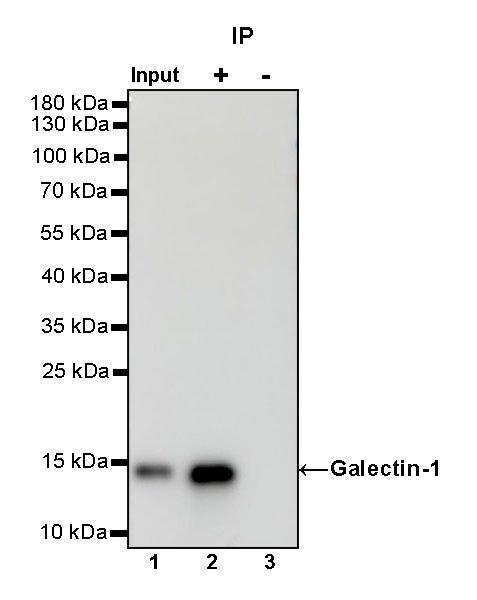
Galectin-1 Rabbit mAb at 1/50 dilution (1 µg) immunoprecipitating Galectin-1 in 0.4 mg HeLa whole cell lysate.
Western blot was performed on the immunoprecipitate using Galectin-1 Rabbit mAb at 1/1000 dilution.
Secondary antibody (HRP) for IP was used at 1/1000 dilution.
Lane 1: HeLa whole cell lysate 20 µg (Input)
Lane 2: Galectin-1 Rabbit mAb IP in HeLa whole cell lysate
Lane 3: Rabbit monoclonal IgG IP in HeLa whole cell lysate
Predicted MW: 15 kDa
Observed MW: 13 kDa
免疫组化
IHC shows positive staining in paraffin-embedded human tonsil. Anti- Galectin-1 antibody was used at 1/1000 dilution, followed by a HRP Polymer for Mouse & Rabbit IgG (ready to use). Counterstained with hematoxylin. Heat mediated antigen retrieval with Tris/EDTA buffer pH9.0 was performed before commencing with IHC staining protocol.
IHC shows positive staining in paraffin-embedded human testis. Anti- Galectin-1 antibody was used at 1/1000 dilution, followed by a HRP Polymer for Mouse & Rabbit IgG (ready to use). Counterstained with hematoxylin. Heat mediated antigen retrieval with Tris/EDTA buffer pH9.0 was performed before commencing with IHC staining protocol.
IHC shows positive staining in paraffin-embedded human kidney. Anti- Galectin-1 antibody was used at 1/1000 dilution, followed by a HRP Polymer for Mouse & Rabbit IgG (ready to use). Counterstained with hematoxylin. Heat mediated antigen retrieval with Tris/EDTA buffer pH9.0 was performed before commencing with IHC staining protocol.
IHC shows positive staining in paraffin-embedded human colon cancer. Anti- Galectin-1 antibody was used at 1/1000 dilution, followed by a HRP Polymer for Mouse & Rabbit IgG (ready to use). Counterstained with hematoxylin. Heat mediated antigen retrieval with Tris/EDTA buffer pH9.0 was performed before commencing with IHC staining protocol.
免疫细胞化学
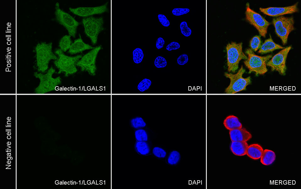
ICC shows positive staining in HeLa cells (top panel) and negative staining in LNCaP cells (below panel). Anti-Galectin-1 antibody was used at 1/500 dilution (Green) and incubated overnight at 4°C. Goat polyclonal Antibody to Rabbit IgG - H&L (Alexa Fluor® 488) was used as secondary antibody at 1/1000 dilution. The cells were fixed with 100% ice-cold methanol and permeabilized with 0.1% PBS-Triton X-100. Nuclei were counterstained with DAPI (Blue). Counterstain with tubulin (Red).


评论(0)