


产品介绍 评论(0)
宿主来源
Mouse分子别名
Integrin alpha-L, CD11 antigen-like family member A, Leukocyte adhesion glycoprotein LFA-1 alpha chain (LFA-1A), Leukocyte function-associated molecule 1 alpha chain, ITGAL细胞定位
Cell membraneAccession
P20701克隆号
S-R402抗体类型
Mouse mAb抗体同种型
IgG1,k应用
FCM, ICC反应种属 ?
Hu纯化方式
Protein G浓度
2 mg/ml标记
Unconjugated性状
Liquid缓冲体系
PBS, 40% Glycerol, 0.05% BSA, 0.03% Proclin 300
储存条件
12 months from date of receipt / reconstitution, -20 °C as supplied.
| 应用 | 稀释度 |
|---|---|
| FCM | 1:200 |
| ICC | 1:50 |
CD11a, also known as Lymphocyte Function-Associated Antigen 1 (LFA-1) alpha chain or integrin alpha L, is an adhesion molecule primarily expressed on leukocytes such as Leu[A5] cells. It plays a crucial role in cellular adhesion by binding to intercellular adhesion molecules (ICAMs) like ICAM-1 (CD54), ICAM-2 (CD102), and ICAM-3 (CD105). Additionally, CD11a can interact with JAM-1, facilitating the passage of leukocytes across endothelial cells. CD11a has various functions in the immune system. For instance, in primary immune thrombocytopenia (ITP), CD11a is expressed on regulatory T cells (Tregs) and interacts with antigen-presenting cells (APCs) to participate in immune regulation. Furthermore, CD11a is associated with the progression of rheumatoid arthritis (RA) and is considered an important factor in the pathogenesis of the disease. In cancer therapy, CD11a also shows promising potential. Researchers have found that activating molecules like CD11a on the surface of macrophages can enhance their ability to kill cancer cells, providing a hopeful approach for cancer treatment.
流式分析
Flow cytometric analysis of human PBMC (human peripheral blood mononuclear cell) labelling Human CD11a antibody at 1/200 (1 μg) / (Red) compared with a Mouse monoclonal IgG (Black) isotype control and an unlabelled control (cells without incubation with primary antibody and secondary antibody) (Blue). Goat Anti - Mouse IgG Alexa Fluor® 488 was used as the secondary antibody. Gated on total viable cells.
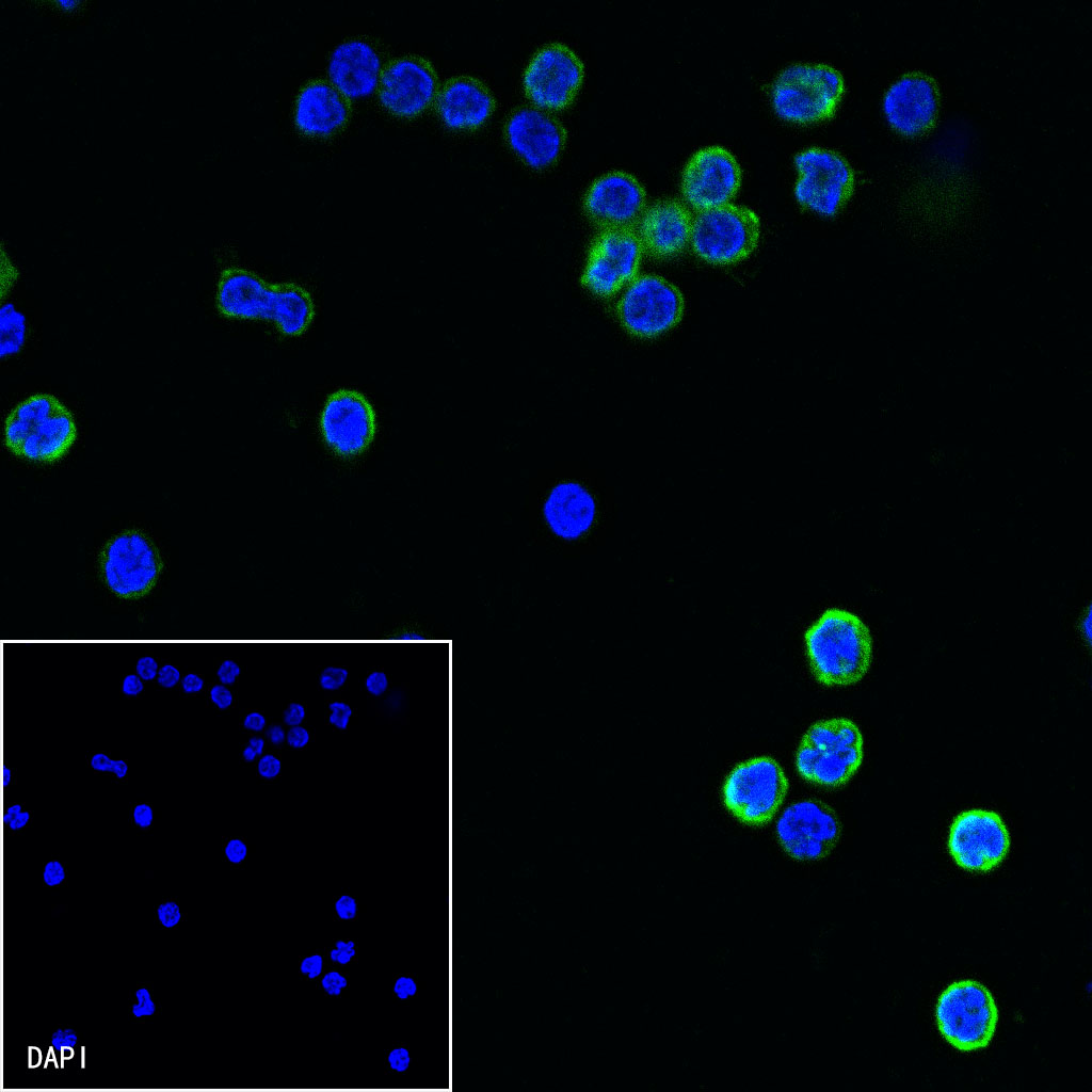
免疫细胞化学
-
ICC shows positive staining in human PBMCs. Anti-CD11a antibody was used at 1/50 dilution (Green) and incubated overnight at 4°C. Goat polyclonal Antibody to Mouse IgG - H&L (Alexa Fluor® 488) was used as secondary antibody at 1/1000 dilution. The cells were fixed with 4% PFA and permeabilized with 0.1% PBS-Triton X-100. Nuclei were counterstained with DAPI (Blue).

评论(0)