




 分享
分享产品介绍 FAQs 评论(0)
宿主来源
Rabbit分子别名
T-lymphocyte activation antigen CD86, Activation B7-2 antigen, B70, BU63, CTLA-4 counter-receptor B7.2, FUN-1免疫原
Recombinant Protein细胞定位
Cell membraneAccession
P42081克隆号
S-990-1抗体类型
Recombinant mAb抗体同种型
IgG反应种属 ?
Hu纯化方式
Protein A浓度
0.5 mg/ml标记
Unconjugated性状
Liquid缓冲体系
PBS, 40% Glycerol, 0.05% BSA, 0.03% Proclin 300
储存条件
12 months from date of receipt / reconstitution, -20 °C as supplied
应用
FCM
ICC ?
WB
IP
稀释度
应用 稀释度 WB 1:1000 ICC 1:500 FCM 1:500 IP 1:50
CD86 protein, also known as B7.2, is a crucial cell surface membrane protein belonging to the immunoglobulin superfamily with a molecular weight of approximately 80Kd. It is highly expressed on the surface of antigen-presenting cells. CD86 is expressed in various immune cells, including dendritic cells, monocytes, memory T lymphocytes, germinal center B lymphocytes, activated B lymphocytes, and activated T lymphocytes. CD86 protein plays a pivotal role in the immune system by interacting with CD28 and CTLA-4 (also known as CD152) proteins on the surface of T cells. The binding of CD86 to CD28 serves as a co-stimulatory signal for T cell activation, while the interaction between CD86 and CTLA-4 negatively regulates T cell activation and reduces immune responses. Additionally, CD86 protein contains two extracellular immunoglobulin domains (IgV and IgC), which bind to intercellular adhesion molecule 1 (ICAM-1) and participate in immune cell contact and signal transduction. Abnormal expression of CD86 is closely associated with the occurrence and development of various immune-related diseases. Therefore, understanding the structure and function of CD86 protein is crucial for revealing its immune regulatory mechanisms and providing new theoretical bases for disease treatment. Furthermore, experimental studies have demonstrated that CD86 protein plays a significant role in the activation and differentiation of immune cells, holding potential therapeutic value in immune-related diseases and offering a new breakthrough in the study of immune regulatory mechanisms.
免疫印迹
WB result of CD86 Rabbit mAb
Primary antibody: CD86 Rabbit mAb at 1/1000 dilution
Lane 1: Jurkat whole cell lysate 20 µg
Lane 2: Raji whole cell lysate 20 µg
Lane 3: Daudi whole cell lysate 20 µg
Lane 4: Ramos whole cell lysate 20 µg
Negative control: Jurkat whole cell lysate
Secondary antibody: Goat Anti-rabbit IgG, (H+L), HRP conjugated at 1/10000 dilution
Predicted MW: 37 kDa
Observed MW: 70~90 kDa
流式分析
Flow cytometric analysis of Jurkat (Human T cell leukemia T lymphocyte, left) / Raji (Human Burkitt's lymphoma B lymphocyte, Right) cells labelling CD86 antibody at 1/500 dilution (0.1 μg) / (Red) compared with a Rabbit monoclonal IgG (Black) isotype control and an unlabelled control (cells without incubation with primary antibody and secondary antibody) (Blue). Goat Anti - Rabbit IgG Alexa Fluor® 488 was used as the secondary antibody.
Negative control: JurkatFlow cytometric analysis of human PBMC (human peripheral blood mononuclear cell) labelling CD86 antibody at 1/500 (0.1 μg) dilution (Right) compared with a Rabbit monoclonal IgG isotype control (Left). Goat Anti - Rabbit IgG Alexa Fluor® 488 was used as the secondary antibody. Then cells were stained with CD14 - Alexa Fluor® 647 separately. Gated on total viable cells.
免疫沉淀
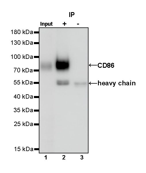
CD86 Rabbit mAb at 1/50 dilution (1 µg) immunoprecipitating CD86 in 0.4 mg Daudi whole cell lysate.
Western blot was performed on the immunoprecipitate using CD86 Rabbit mAb at 1/1000 dilution.
Secondary antibody (HRP) for IP was used at 1/1000 dilution.
Lane 1: Daudi whole cell lysate 20 µg (Input)
Lane 2: CD86 Rabbit mAb IP in Daudi whole cell lysate
Lane 3: Rabbit monoclonal IgG IP in Daudi whole cell lysate
Predicted MW: 37 kDa
Observed MW: 70~90 kDa
免疫细胞化学
ICC shows positive staining in Raji cells (top panel) and negative staining in Jurkat cells (below panel). Anti-CD86 antibody was used at 1/500 dilution (Green) and incubated overnight at 4°C. Goat polyclonal Antibody to Rabbit IgG - H&L (Alexa Fluor® 488) was used as secondary antibody at 1/1000 dilution. The cells were fixed with 4% PFA and permeabilized with 0.1% PBS-Triton X-100. Nuclei were counterstained with DAPI (Blue). Counterstain with tubulin (Red).
FAQs
我们一般不推荐客户回收利用抗体。 因为抗体使用之后缓冲体系已经发生改变,不同客户在回收抗体的保存条件上也会有差异,所以抗体回收使用效果无法保证。另外,我们对一批抗体回收验证测试,测试结果显示不同抗体可回收次数不同,一般效价越高的抗体,可重复使用的次数越多,客户可根据实验情况来确定

评论(0)