


 申请试用
申请试用 产品介绍 FAQs 评论(0)
宿主来源
Rabbit抗原名称
Phospho-Akt (Ser473)分子别名
RAC-alpha serine/threonine-protein kinase, Protein kinase B (PKB), Protein kinase B alpha (PKB alpha), Proto-oncogene c-Akt, RAC-PK-alpha, PKB, RAC, AKT1免疫原
Synthetic Peptide细胞定位
Cytoplasm, Nucleus, Cell membraneAccession
P31749克隆号
S-622-64抗体类型
Recombinant mAb抗体同种型
IgG翻译后修饰类型
磷酸化反应种属 ?
Hu预测反应种属
(反应种属缩写表)Rt, Ms纯化方式
Protein A浓度
0.5 mg/ml标记
Unconjugated性状
Liquid缓冲体系
PBS, 40% Glycerol, 0.05% BSA, 0.03% Proclin 300储存条件
12 months from date of receipt / reconstitution, -20 °C as supplied
应用
ICFCM ,ICC ? ,WB ,IP稀释度
应用 稀释度 WB 1:1000 ICC 1:500 ICFCM 1:500 Dot Blot 1:1000 IP 1:50
Akt or Protein kinase B, is a serine/threonine kinase that plays an important role in regulating a number of cellular processes such as growth, metabolism and survival. The importance of the Akt pathway is highlighted by the mutation of various components of the pathway in human cancers such as the PTEN and PI3-kinase (P110α), which occur in more than 30% of human tumors. For Akt to achieve full activation, phosphorylation is needed at both serine 473 (ser473) of the hydrophobic tail and threonine 308 (thr308) of the activation motif, upon growth factor ligation to the receptor tyrosine kinases.
免疫印迹
WB result of Phospho-Akt (Ser473) Rabbit mAb
Primary antibody: Phospho-Akt (Ser473) Rabbit mAb at 1/1000 dilution
Lane 1: untreated Jurkat whole cell lysate 20 µg
Lane 2: Jurkat treated with 100 nM Calyculin A for 30 minutes whole cell lysate 20 µg
Secondary antibody: Goat Anti-rabbit IgG, (H+L), HRP conjugated at 1/10000 dilution
Predicted MW: 56 kDa
Observed MW: 60 kDa
流式分析
Flow cytometric analysis of 4% PFA fixed 90% methanol permeabilized Jurkat (Human T cell leukemia T lymphocyte) cells, treated with 100nM Calyculin A for 30 min (Red) or untreated (Green), labeling Phospho-Akt (Ser473) at 1/500 dilution (0.1 μg) compared with a rabbit monoclonal IgG isotype control (Black) and an unlabeled control (cells without incubation with primary antibody and secondary antibody) (Blue). Goat Anti - Rabbit IgG Alexa Fluor® 488 was used as the secondary antibody.
免疫沉淀
Phospho-Akt (Ser473) Rabbit mAb at 1/50 dilution (1 µg) immunoprecipitating Phospho-Akt (Ser473) in 0.4 mg Jurkat treated with 100 nM Calyculin A for 30 minutes whole cell lysate.
Western blot was performed on the immunoprecipitate using Phospho-Akt (Ser473) Rabbit mAb at 1/1000 dilution.
Secondary antibody (HRP) for IP was used at 1/1000 dilution.
Lane 1: Jurkat treated with 100 nM Calyculin A for 30 minutes whole cell lysate 20 µg (Input)
Lane 2: Phospho-Akt (Ser473) Rabbit mAb IP in Jurkat treated with 100 nM Calyculin A for 30 minutes whole cell lysate
Lane 3: Rabbit monoclonal IgG IP in Jurkat treated with 100 nM Calyculin A for 30 minutes whole cell lysate
Predicted MW: 56 kDa
Observed MW: 60 kDa
斑点杂交
Dot blot result of Phospho-Akt (Ser473) Rabbit mAb
Lane1: Phospho-Akt (Ser473) peptide
Lane2: Akt unmodified peptide Primary antibody: Phospho-Akt (Ser473) Rabbit mAb at 1/1000 dilution
Secondary antibody: Goat Anti-Rabbit IgG, (H+L), HRP conjugated at 1/10000 dilution
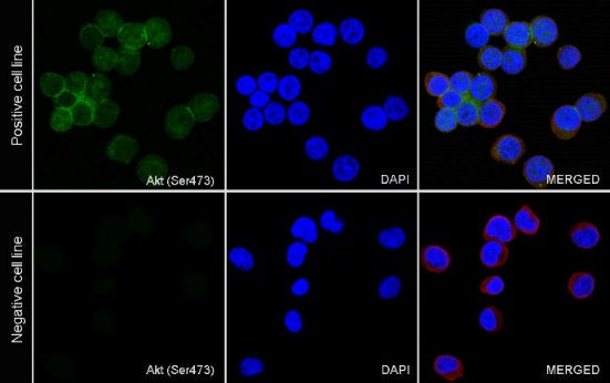
免疫细胞化学
ICC analysis of Jurkat cells treated with Calyculin A (100nM, 30 min) (top panel) and Jurkat cells untreated with Calyculin A (100nM, 30 min) (below panel). Anti-Phospho-Akt (Ser473) antibody was used at 1/500 dilution (Green) and incubated overnight at 4°C. Goat polyclonal Antibody to Rabbit IgG - H&L (Alexa Fluor® 488) was used as secondary antibody at 1/1000 dilution. The cells were fixed with 4% PFA and permeabilized with 0.1% PBS-Triton X-100. Nuclei were counterstained with DAPI (Blue). Counterstain with tubulin (Red).
FAQs
我们一般不推荐客户回收利用抗体。 因为抗体使用之后缓冲体系已经发生改变,不同客户在回收抗体的保存条件上也会有差异,所以抗体回收使用效果无法保证。另外,我们对一批抗体回收验证测试,测试结果显示不同抗体可回收次数不同,一般效价越高的抗体,可重复使用的次数越多,客户可根据实验情况来确定
我们推荐客户使用TPST+5%脱脂奶粉来稀释一抗,进行封闭。 虽然BSA被推荐为WB检测磷酸化蛋白的常用封闭剂,但是脱脂奶粉获取更加方便,覆盖更广泛的非特异性结合位点,在一抗性能优越的前提下,使用脱脂奶粉封闭性价比更高

评论(0)