


 大包装询价
大包装询价 产品介绍 评论(0)
宿主来源
Rabbit分子别名
Protein kinase C alpha type, PKC-A, PKC-alpha, PRKCA, PKCA, PRKACA免疫原
Synthetic Peptide细胞定位
Nucleus, Cell membrane, CytoplasmAccession
P17252克隆号
S-624-36抗体类型
Recombinant mAb抗体同种型
IgG应用
ICFCM, IHC-P, ICC, WB, IP反应种属 ?
Hu, Ms, Rt预测反应种属
(反应种属缩写表)Bv纯化方式
Protein A浓度
0.5 mg/ml标记
Unconjugated性状
Liquid缓冲体系
PBS, 40% Glycerol, 0.05% BSA, 0.03% Proclin 300储存条件
12 months from date of receipt / reconstitution, -20 °C as supplied
| 应用 | 稀释度 |
|---|---|
| WB | 1:1000 |
| IP | 1:50 |
| IHC-P | 1:500 |
| ICFCM | 1:500 |
| ICC | 1:500 |
PKC alpha is one of the PKC family members. This kinase has been reported to play roles in many different cellular processes, such as cell adhesion, cell transformation, cell cycle checkpoint, and cell volume control. Knockout studies in mice suggest that this kinase may be a fundamental regulator of cardiac contractility and Ca2+ handling in myocytes. Increased activation of PKCα is associated with the growth and invasion of cancers. High levels of PKCα are linked to malignant brain cancer. Moreover, a high proliferation rate of glioma tumor cells is the result of overexpression of isozyme PKCα.
免疫印迹
WB result of PKC alpha Rabbit mAb
Primary antibody: PKC alpha Rabbit mAb at 1/1000 dilution
Lane 1: HeLa whole cell lysate 20 µg
Lane 2: SH-SY5Y whole cell lysate 20 µg
Secondary antibody: Goat Anti-Rabbit IgG, (H+L), HRP conjugated at 1/10000 dilution
Predicted MW: 77 kDa
Observed MW: 85 kDaWB result of PKC alpha Rabbit mAb
Primary antibody: PKC alpha Rabbit mAb at 1/1000 dilution
Lane 1: NIH/3T3 whole cell lysate 20 µg
Lane 2: mouse brain lysate 20 µg
Lane 3: mouse spleen lysate 20 µg
Secondary antibody: Goat Anti-Rabbit IgG, (H+L), HRP conjugated at 1/10000 dilution
Predicted MW: 77 kDa
Observed MW: 85 kDaWB result of PKC alpha Rabbit mAb
Primary antibody: PKC alpha Rabbit mAb at 1/1000 dilution
Lane 1: C6 whole cell lysate 20 µg
Lane 2: rat brain lysate 20 µg
Secondary antibody: Goat Anti-Rabbit IgG, (H+L), HRP conjugated at 1/10000 dilution
Predicted MW: 77 kDa
Observed MW: 85 kDa
流式分析
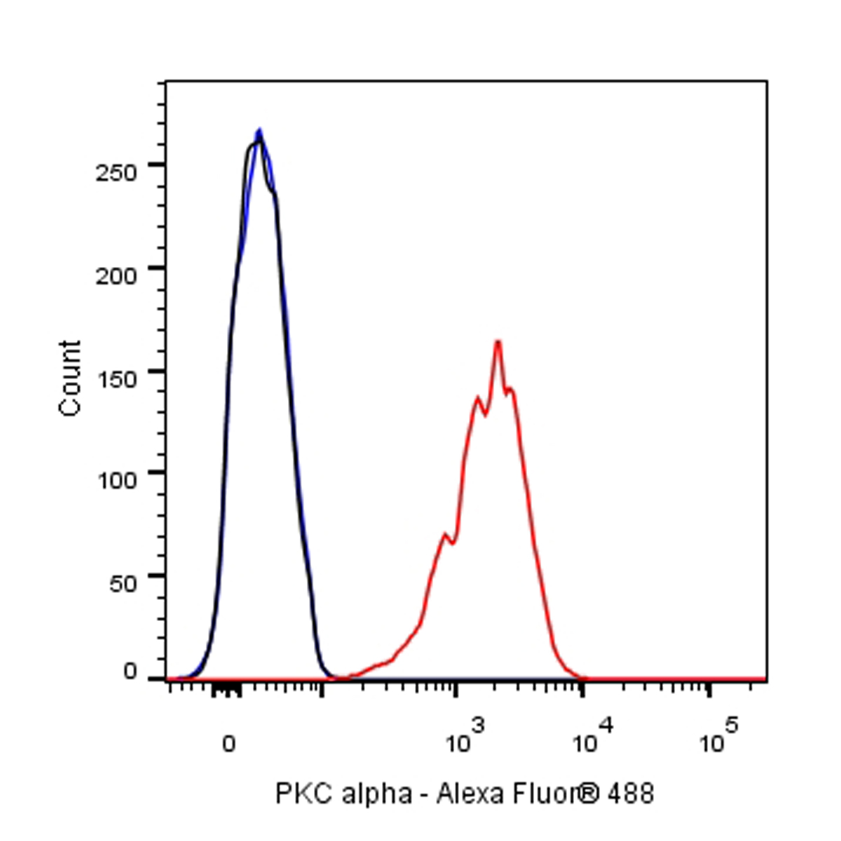
Flow cytometric analysis of 4% PFA fixed 90% methanol permeabilized SH-SY5Y (Human neuroblastoma epithelial cell) labelling PKC alpha antibody at 1/500 dilution (0.1 μg)/ (Right) compared with a Rabbit monoclonal IgG / (Left) isotype control. Goat Anti - Rabbit IgG Alexa Fluor® 488 was used as the secondary antibody.
免疫沉淀
PKC alpha Rabbit mAb at 1/50 dilution (1 µg) immunoprecipitating PKC alpha in 0.4 mg SH-SY5Y whole cell lysate.
Western blot was performed on the immunoprecipitate using PKC alpha Rabbit mAb at 1/1000 dilution.
Secondary antibody (HRP) for IP was used at 1/400 dilution.
Lane 1: SH-SY5Y whole cell lysate 20 µg (Input)
Lane 2: PKC alpha Rabbit mAb IP in SH-SY5Y whole cell lysate
Lane 3: Rabbit monoclonal IgG IP in SH-SY5Y whole cell lysate
Predicted MW: 77 kDa
Observed MW: 85 kDa
免疫组化
IHC shows positive staining in paraffin-embedded human cerebral cortex. Anti-PKC alpha antibody was used at 1/500 dilution, followed by a HRP Polymer for Mouse & Rabbit IgG (ready to use). Counterstained with hematoxylin. Heat mediated antigen retrieval with Tris/EDTA buffer pH9.0 was performed before commencing with IHC staining protocol.
IHC shows positive staining in paraffin-embedded human breast cancer. Anti-PKC alpha antibody was used at 1/500 dilution, followed by a HRP Polymer for Mouse & Rabbit IgG (ready to use). Counterstained with hematoxylin. Heat mediated antigen retrieval with Tris/EDTA buffer pH9.0 was performed before commencing with IHC staining protocol.
IHC shows positive staining in paraffin-embedded human gastric cancer. Anti-PKC alpha antibody was used at 1/500 dilution, followed by a HRP Polymer for Mouse & Rabbit IgG (ready to use). Counterstained with hematoxylin. Heat mediated antigen retrieval with Tris/EDTA buffer pH9.0 was performed before commencing with IHC staining protocol.
IHC shows positive staining in paraffin-embedded human pancreatic cancer. Anti-PKC alpha antibody was used at 1/500 dilution, followed by a HRP Polymer for Mouse & Rabbit IgG (ready to use). Counterstained with hematoxylin. Heat mediated antigen retrieval with Tris/EDTA buffer pH9.0 was performed before commencing with IHC staining protocol.
IHC shows positive staining in paraffin-embedded mouse cerebral cortex. Anti-PKC alpha antibody was used at 1/500 dilution, followed by a HRP Polymer for Mouse & Rabbit IgG (ready to use). Counterstained with hematoxylin. Heat mediated antigen retrieval with Tris/EDTA buffer pH9.0 was performed before commencing with IHC staining protocol.
IHC shows positive staining in paraffin-embedded rat cerebral cortex. Anti-PKC alpha antibody was used at 1/500 dilution, followed by a HRP Polymer for Mouse & Rabbit IgG (ready to use). Counterstained with hematoxylin. Heat mediated antigen retrieval with Tris/EDTA buffer pH9.0 was performed before commencing with IHC staining protocol.
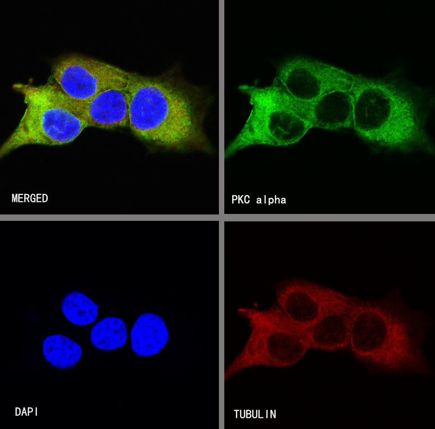
免疫细胞化学

ICC shows positive staining in SH-SY5Y cells. Anti-PKC alpha antibody was used at 1/500 dilution (Green) and incubated overnight at 4°C. Goat polyclonal Antibody to Rabbit IgG - H&L (Alexa Fluor® 488) was used as secondary antibody at 1/1000 dilution. The cells were fixed with 100% ice-cold methanol and permeabilized with 0.1% PBS-Triton X-100. Nuclei were counterstained with DAPI (Blue).Counterstain with tubulin (red).
ICC shows positive staining in SH-SY5Y cells. Anti-PKC alpha antibody was used at 1/500 dilution (Green) and incubated overnight at 4°C. Goat polyclonal Antibody to Rabbit IgG - H&L (Alexa Fluor® 488) was used as secondary antibody at 1/1000 dilution. The cells were fixed with 100% ice-cold methanol and permeabilized with 0.1% PBS-Triton X-100. Nuclei were counterstained with DAPI (Blue).Counterstain with tubulin (red).

评论(0)