


 大包装询价
大包装询价 产品介绍 评论(0)
宿主来源
Rabbit抗原名称
Fer分子别名
Tyrosine-protein kinase Fer, Feline encephalitis virus-related kinase FER, Fujinami poultry sarcoma/Feline sarcoma-related protein Fer, Proto-oncogene c-Fer, Tyrosine kinase 3, p94-Fer, TYK3细胞定位
Nucleus, Cell membrane, CytoplasmAccession
P16591克隆号
S-R303抗体类型
Recombinant mAb抗体同种型
IgG应用
ICFCM, ICC, WB反应种属 ?
Hu, Ms, Rt纯化方式
Protein A浓度
0.5 mg/ml标记
Unconjugated性状
Liquid缓冲体系
PBS, 40% Glycerol, 0.05%BSA, 0.03% Proclin 300储存条件
12 months from date of receipt / reconstitution, -20 °C as supplied
| 应用 | 稀释度 |
|---|---|
| WB | 1:1000 |
| ICFCM | 1:50 |
| ICC | 1:500 |
Fer protein is a member of the FPS/FES family of nontransmembrane receptor tyrosine kinases. It regulates cell-cell adhesion and mediates signaling from the cell surface to the cytoskeleton via growth factor receptors.
免疫印迹
WB result of Fer Rabbit mAb
Primary antibody: Fer Rabbit mAb at 1/1000 dilution
Lane 1: HeLa whole cell lysate 20 µg
Lane 2: Jurkat whole cell lysate 20 µg
Lane 3: A549 whole cell lysate 20 µg
Lane 4: LNCaP whole cell lysate 20 µg
Secondary antibody: Goat Anti-Rabbit IgG, (H+L), HRP conjugated at 1/10000 dilution
Predicted MW: 95 kDa
Observed MW: 95 kDa
(This blot was developed with high sensitivity substrate)WB result of Fer Rabbit mAb
Primary antibody: Fer Rabbit mAb at 1/1000 dilution
Lane 1: PC-12 whole cell lysate 20 µg
Secondary antibody: Goat Anti-Rabbit IgG, (H+L), HRP conjugated at 1/10000 dilution
Predicted MW: 95 kDa
Observed MW: 95 kDa
WB result of Fer Rabbit mAb
Primary antibody: Fer Rabbit mAb at 1/1000 dilution
Lane 1: NIH/3T3 whole cell lysate 5 µg
Secondary antibody: Goat Anti-Rabbit IgG, (H+L), HRP conjugated at 1/10000 dilution
Predicted MW: 95 kDa
Observed MW: 95 kDa
流式分析

Flow cytometric analysis of 4% PFA fixed 90% methanol permeabilized HeLa (Human cervix adenocarcinoma epithelial cell) cells labelling Fer antibody at 1/50 dilution (1 μg)/ (Red) compared with a Rabbit monoclonal IgG (Black) isotype control and an unlabelled control (cells without incubation with primary antibody and secondary antibody) (Blue). Goat Anti - Rabbit IgG Alexa Fluor® 488 was used as the secondary antibody.
免疫细胞化学
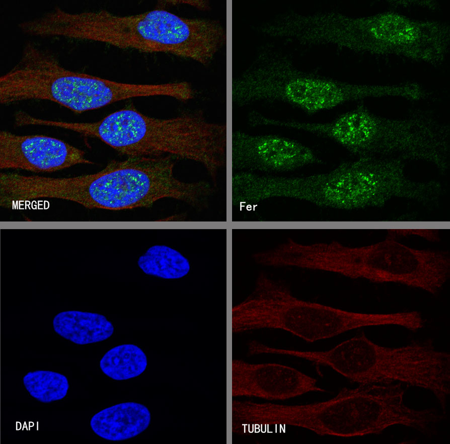
ICC shows positive staining in HeLa cells. Anti-Fer antibody was used at 1/500 dilution (Green) and incubated overnight at 4°C. Goat polyclonal Antibody to Rabbit IgG - H&L (Alexa Fluor® 488) was used as secondary antibody at 1/1000 dilution. The cells were fixed with 100% ice-cold methanol and permeabilized with 0.1% PBS-Triton X-100. Nuclei were counterstained with DAPI (Blue).Counterstain with tubulin (red).

评论(0)