


 大包装询价
大包装询价 产品介绍 评论(0)
宿主来源
Rabbit抗原名称
TFEB分子别名
Transcription factor EB, Class E basic helix-loop-helix protein 35 (bHLHe35), BHLHE35细胞定位
Cytoplasm, NucleusAccession
P19484克隆号
S-R296抗体类型
Recombinant mAb抗体同种型
IgG应用
ICFCM, IHC-P, ICC, WB, IP反应种属 ?
Hu纯化方式
Protein A浓度
0.5 mg/ml标记
Unconjugated性状
Liquid缓冲体系
PBS, 40% Glycerol, 0.05%BSA, 0.03% Proclin 300储存条件
12 months from date of receipt / reconstitution, -20 °C as supplied
| 应用 | 稀释度 |
|---|---|
| WB | 1:1000 |
| IHC | 1:500 |
| ICFCM | 1:500 |
| ICC | 1:500 |
| IP | 1:50 |
TFEB is a master gene for lysosomal biogenesis. It encodes a transcription factor that coordinates expression of lysosomal hydrolases, membrane proteins and genes involved in autophagy. Upon nutrient depletion and under aberrant lysosomal storage conditions such as in lysosomal storage diseases, TFEB translocates from the cytoplasm to the nucleus, resulting in the activation of its target genes. TFEB overexpression in cultured cells induces lysosomal biogenesis, exocytosis and autophagy. TFEB constitutive activation, due to FLCN mutations, drives renal cystogenesis and tumorigenesis in Birt–Hogg–Dubé syndrome.
免疫印迹
WB result of TFEB Rabbit mAb
Primary antibody: TFEB Rabbit mAb at 1/1000 dilution
Lane 1: A549 whole cell lysate 20 µg
Lane 2: Raji whole cell lysate 20 µg
Negative control: A549 whole cell lysate
Secondary antibody: Goat Anti-Rabbit IgG, (H+L), HRP conjugated at 1/10000 dilution
Predicted MW: 53 kDa
Observed MW: 66 kDa
(This blot was developed with high sensitivity substrate)
流式分析
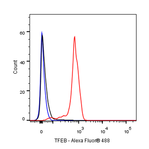
Flow cytometric analysis of 4% PFA fixed 90% methanol permeabilized Raji (Human Burkitt's lymphoma B lymphocyte) cells labelling TFEB antibody at 1/500 dilution (0.1 μg)/ (Red) compared with a Rabbit monoclonal IgG (Black) isotype control and an unlabelled control (cells without incubation with primary antibody and secondary antibody) (Blue). Goat Anti - Rabbit IgG Alexa Fluor® 488 was used as the secondary antibody.
免疫沉淀
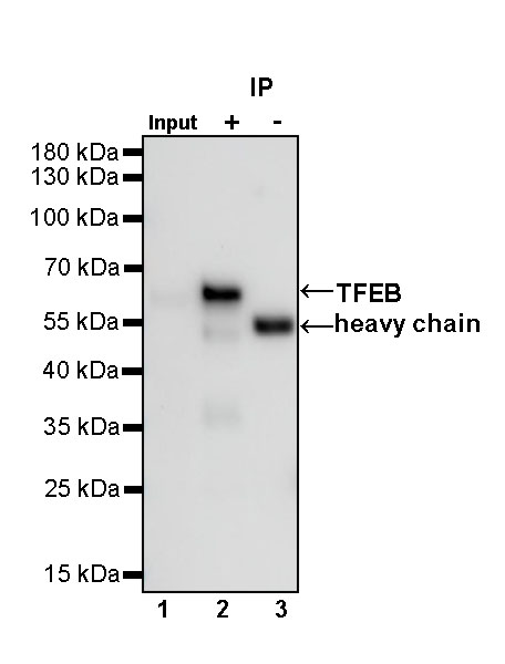
TFEB Rabbit mAb at 1/50 dilution (1 µg) immunoprecipitating TFEB in 0.4 mg Raji whole cell lysate.
Western blot was performed on the immunoprecipitate using TFEB Rabbit mAb at 1/1000 dilution.
Secondary antibody (HRP) for IP was used at 1/400 dilution.
Lane 1: Raji whole cell lysate 20 µg (Input)
Lane 2: TFEB Rabbit mAb IP in Raji whole cell lysate
Lane 3: Rabbit monoclonal IgG IP in Raji whole cell lysate
Predicted MW: 53 kDa
Observed MW: 66 kDa
免疫组化
IHC shows positive staining in paraffin-embedded human breast. Anti-TFEB antibody was used at 1/500 dilution, followed by a HRP Polymer for Mouse & Rabbit IgG (ready to use). Counterstained with hematoxylin. Heat mediated antigen retrieval with Tris/EDTA buffer pH9.0 was performed before commencing with IHC staining protocol.
IHC shows positive staining in paraffin-embedded human colon. Anti-TFEB antibody was used at 1/500 dilution, followed by a HRP Polymer for Mouse & Rabbit IgG (ready to use). Counterstained with hematoxylin. Heat mediated antigen retrieval with Tris/EDTA buffer pH9.0 was performed before commencing with IHC staining protocol.
IHC shows positive staining in paraffin-embedded human kidney. Anti-TFEB antibody was used at 1/500 dilution, followed by a HRP Polymer for Mouse & Rabbit IgG (ready to use). Counterstained with hematoxylin. Heat mediated antigen retrieval with Tris/EDTA buffer pH9.0 was performed before commencing with IHC staining protocol.
IHC shows positive staining in paraffin-embedded human breast cancer. Anti-TFEB antibody was used at 1/500 dilution, followed by a HRP Polymer for Mouse & Rabbit IgG (ready to use). Counterstained with hematoxylin. Heat mediated antigen retrieval with Tris/EDTA buffer pH9.0 was performed before commencing with IHC staining protocol.
IHC shows positive staining in paraffin-embedded human endometrial cancer. Anti-TFEB antibody was used at 1/500 dilution, followed by a HRP Polymer for Mouse & Rabbit IgG (ready to use). Counterstained with hematoxylin. Heat mediated antigen retrieval with Tris/EDTA buffer pH9.0 was performed before commencing with IHC staining protocol.
免疫细胞化学
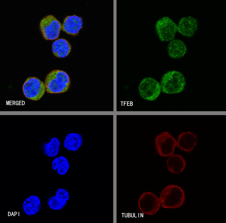
ICC shows positive staining in Raji cells. Anti-TFEB antibody was used at 1/500 dilution (Green) and incubated overnight at 4°C. Goat polyclonal Antibody to Rabbit IgG - H&L (Alexa Fluor® 488) was used as secondary antibody at 1/1000 dilution. The cells were fixed with 4%PFA and permeabilized with 0.1% PBS-Triton X-100. Nuclei were counterstained with DAPI (Blue).Counterstain with tubulin (red).

评论(0)