




 分享
分享产品介绍 FAQs 评论(0)
宿主来源
Mouse抗原名称
CD2分子别名
T-cell surface antigen CD2, Erythrocyte receptor, LFA-2, LFA-3 receptor, Rosette receptor, T-cell surface antigen T11/Leu-5, SRBC免疫原
Recombinant Protein细胞定位
Cell membraneAccession
P06729克隆号
S-633-25抗体类型
Mouse mAb抗体同种型
IgG1,k反应种属 ?
Hu纯化方式
Protein G浓度
2 mg/ml标记
Unconjugated性状
Liquid缓冲体系
PBS, 40% Glycerol, 0.05%BSA, 0.03% Proclin 300储存条件
12 months from date of receipt / reconstitution, -20 °C as supplied
应用
FCM
ICC ?
稀释度
应用 稀释度 FCM 1:2000 ICC 1:500
CD2 (cluster of differentiation 2) is a cell adhesion molecule found on the surface of T cells and natural killer (NK) cells. It has also been called T-cell surface antigen T11/Leu-5, LFA-2, LFA-3 receptor, erythrocyte receptor and rosette receptor. It interacts with other adhesion molecules, such as lymphocyte function-associated antigen-3 (LFA-3/CD58) in humans, or CD48 in rodents, which are expressed on the surfaces of other cells. CD2 is a specific marker for T cells and NK cells, and can therefore be used in immunohistochemistry to identify the presence of such cells in tissue sections. The great majority of T cell lymphomas and leukaemias also express CD2, making it possible to use the presence of the antigen to distinguish these conditions from B cell neoplasms.
流式分析
Flow cytometric analysis of Ramos (Human Burkitt's lymphoma B lymphocyte, left) / Jurkat (Human T cell leukemia T lymphocyte, right) cells labelling CD2 antibody at 1/2000 dilution (0.1 μg) / (red) compared with a Mouse monoclonal IgG (Black) isotype control and an unlabelled control (cells without incubation with primary antibody and secondary antibody) (Blue). Goat Anti - Mouse IgG Alexa Fluor® 488 was used as the secondary antibody.
免疫细胞化学
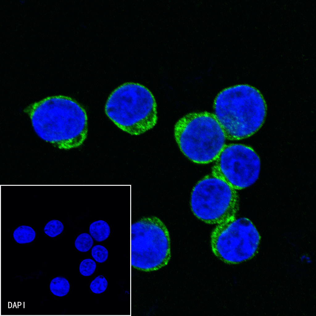
ICC shows positive staining in Jurkat cells. Anti-CD2 antibody was used at 1/500 dilution (Green) and incubated overnight at 4°C. Goat polyclonal Antibody to Mouse IgG - H&L (Alexa Fluor® 488) was used as secondary antibody at 1/1000 dilution. The cells were fixed with 4%PFA and permeabilized with 0.1% PBS-Triton X-100. Nuclei were counterstained with DAPI (Blue).
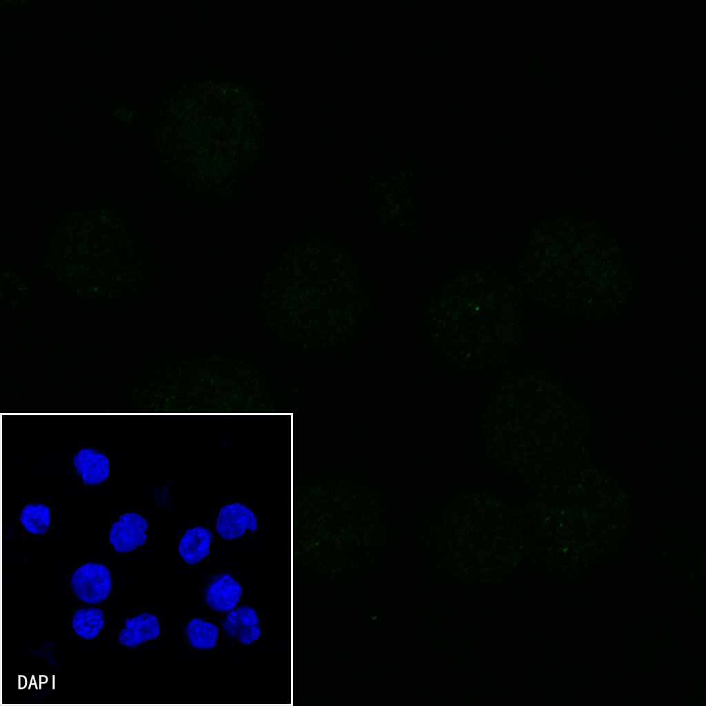
Negative control: ICC shows negative staining in Daudi cells. Anti-CD2 antibody was used at 1/500 dilution and incubated overnight at 4°C. Goat polyclonal Antibody to Mouse IgG - H&L (Alexa Fluor® 488) was used as secondary antibody at 1/1000 dilution. The cells were fixed with 4%PFA and permeabilized with 0.1% PBS-Triton X-100. Nuclei were counterstained with DAPI (Blue).
FAQs
我们一般不推荐客户回收利用抗体。 因为抗体使用之后缓冲体系已经发生改变,不同客户在回收抗体的保存条件上也会有差异,所以抗体回收使用效果无法保证。另外,我们对一批抗体回收验证测试,测试结果显示不同抗体可回收次数不同,一般效价越高的抗体,可重复使用的次数越多,客户可根据实验情况来确定

评论(0)