


 大包装询价
大包装询价 产品介绍 评论(0)
宿主来源
Rabbit抗原名称
Neurofilament M分子别名
Neurofilament medium polypeptide, NF-M, 160 kDa neurofilament protein, Neurofilament 3, Neurofilament triplet M protein, NEFM, NEF3, NFM免疫原
Synthetic Peptide细胞定位
Cytoplasm, CytoskeletonAccession
P07197克隆号
S-501-31抗体类型
Recombinant mAb应用
IHC-P, WB, IP, IF反应种属 ?
Hu, Ms, Rt预测反应种属
(反应种属缩写表)Bv纯化方式
Protein A浓度
0.5 mg/ml标记
Unconjugated性状
Liquid缓冲体系
PBS, 40% Glycerol, 0.05%BSA, 0.03% Proclin 300
储存条件
12 months from date of receipt / reconstitution, -20 °C as supplied
| 应用 | 稀释度 |
|---|---|
| WB | 1:1000 |
| IHC | 1:500 |
| IP | 1:50 |
| IF | 1:2000 |
Neurofilament medium polypeptide (NF-M) is a protein that in humans is encoded by the NEFM gene. Neurofilaments are type IV intermediate filament heteropolymers composed of light (NEFL), medium (this protein), and heavy (NEFH) chains. Neurofilaments comprise the exoskeleton and functionally maintain neuronal caliber. They may also play a role in intracellular transport to axons and dendrites. This gene encodes the medium neurofilament protein. This protein is commonly used as a biomarker of neuronal damage.
免疫印迹
WB result of Neurofilament M Rabbit mAb
Primary antibody: Neurofilament M Rabbit mAb at 1/1000 dilution
Lane 1: mouse liver lysate 20 µg
Lane 2: mouse cerebellum lysate 20 µg
Negative control: mouse liver lysate
Secondary antibody: Goat Anti-Rabbit IgG, (H+L), HRP conjugated at 1/10000 dilution
Predicted MW: 102 kDa
Observed MW: 160 kDaWB result of Neurofilament M Rabbit mAb
Primary antibody: Neurofilament M Rabbit mAb at 1/1000 dilution
Lane 1: rat liver lysate 20 µg
Lane 2: rat brain lysate 20 µg
Negative control: rat liver lysate
Secondary antibody: Goat Anti-Rabbit IgG, (H+L), HRP conjugated at 1/10000 dilution
Predicted MW: 102 kDa
Observed MW: 160 kDa
免疫沉淀
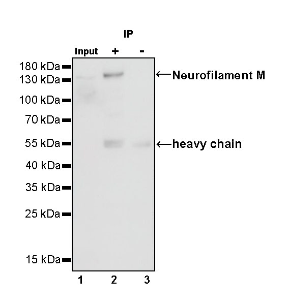
Neurofilament M Rabbit mAb at 1/50 dilution (1 µg) immunoprecipitating Neurofilament M in 0.4 mg mouse cerebellum lysate.
Western blot was performed on the immunoprecipitate using Neurofilament M Rabbit mAb at 1/1000 dilution.
Secondary antibody (HRP) for IP was used at 1/400 dilution.
Lane 1: mouse cerebellum lysate 20 µg (Input)
Lane 2: Neurofilament M Rabbit mAb IP in mouse cerebellum lysate
Lane 3: Rabbit monoclonal IgG IP in mouse cerebellum lysate
Predicted MW: 102 kDa
Observed MW: 160 kDa
免疫组化
IHC shows positive staining in paraffin-embedded human cerebral cortex. Anti- Neurofilament M antibody was used at 1/500 dilution, followed by a HRP Polymer for Mouse & Rabbit IgG (ready to use). Counterstained with hematoxylin. Heat mediated antigen retrieval with Tris/EDTA buffer pH9.0 was performed before commencing with IHC staining protocol.
IHC shows positive staining in paraffin-embedded human cerebellum. Anti- Neurofilament M antibody was used at 1/500 dilution, followed by a HRP Polymer for Mouse & Rabbit IgG (ready to use). Counterstained with hematoxylin. Heat mediated antigen retrieval with Tris/EDTA buffer pH9.0 was performed before commencing with IHC staining protocol.
Negative control: IHC shows negative staining in paraffin-embedded human liver. Anti- Neurofilament M antibody was used at 1/500 dilution, followed by a HRP Polymer for Mouse & Rabbit IgG (ready to use). Counterstained with hematoxylin. Heat mediated antigen retrieval with Tris/EDTA buffer pH9.0 was performed before commencing with IHC staining protocol.
免疫荧光
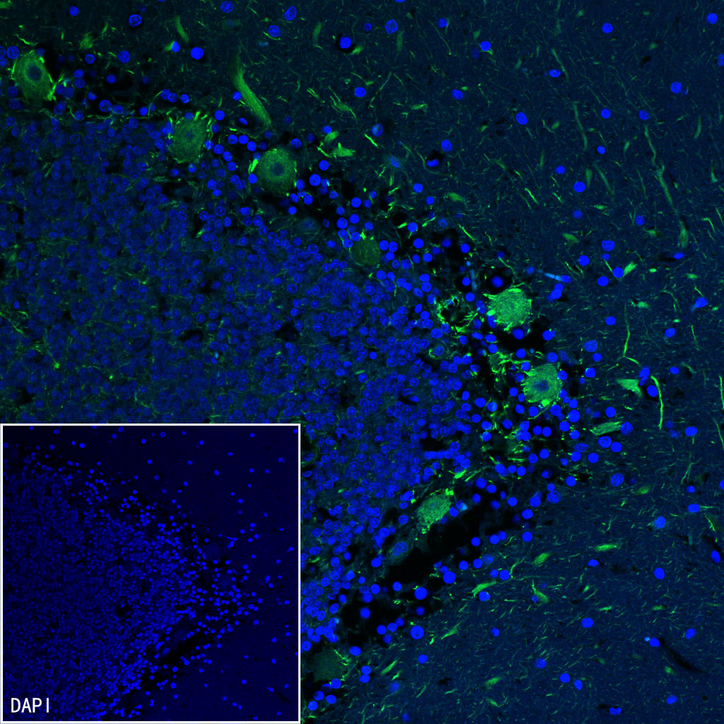
IF shows positive staining in paraffin-embedded human cerebellum. Anti-Neurofilament M antibody was used at 1/2000 dilution (Green) and incubated overnight at 4°C. Goat polyclonal Antibody to Rabbit IgG - H&L (Alexa Fluor® 488) was used as secondary antibody at 1/1000 dilution. Counterstained with DAPI (Blue). Heat mediated antigen retrieval with EDTA buffer pH9.0 was performed before commencing with IF staining protocol.

评论(0)