


 申请试用
申请试用 产品介绍 FAQs 评论(0)
宿主来源
Rabbit抗原名称
FOXA2分子别名
Hepatocyte nuclear factor 3-beta, HNF-3-beta, HNF-3B, Forkhead box protein A2, TCF-3B免疫原
N/A细胞定位
Cytoplasm, NucleusAccession
Q9Y261克隆号
SDT-R174抗体类型
Recombinant mAb反应种属 ?
Hu, Ms, Rt纯化方式
Protein A浓度
0.5 mg/ml标记
Unconjugated性状
Liquid缓冲体系
PBS, 40% Glycerol, 0.05% BSA, 0.03% Proclin 300
储存条件
12 months from date of receipt / reconstitution, -20 °C as supplied
应用
ICFCM ,ChIP ,IHC-P ? ,ICC ? ,WB ,IP稀释度
应用 稀释度 推荐种属 WB 1:1000 Hu IHC-P 1:500 Hu, Ms, Rt ICC 1:200-1:500 Hu IP 1:50 Hu ChIP 1:20-1:50 Hu
FOXA2 is member of a large family of nuclear transcription factors, termed the winged helix family of transcription factors, that are involved in cell commitment, differentiation, and gene transcription in a variety of organs, such as the central nervous system and derivatives of the foregut endoderm, including the gastrointestinal tract, lung, and liver. FOXA2 is required for the formation of foregut endoderm, from which the lung bud is derived, and plays a critical role in organogenesis of the lung.
免疫印迹
WB result of FOXA2 Rabbit mAb
Primary antibody: FOXA2 Rabbit mAb at 1/1000 dilution
Lane 1: LNCaP whole cell lysate 20 µg
Lane 2: PC-3 whole cell lysate 20 µg
Lane 3: SW480 whole cell lysate 20 µg
Lane 4: HepG2 whole cell lysate 20 µg
Negative control: LNCaP whole cell lysate
Secondary antibody: Goat Anti-Rabbit IgG, (H+L), HRP conjugated at 1/10000 dilution
Predicted MW: 48kDa
Observed MW: 48kDa
流式分析
Flow cytometric analysis of HeLa (Human cervix adenocarcinoma epithelial cell, left) / HepG2 (Human hepatocellular carcinoma epithelial cell, right) cells labelling FOXA2 antibody at 1/500 dilution (0.1 μg)/ (red) compared with a Rabbit monoclonal IgG (Black) isotype control and an unlabelled control (cells without incubation with primary antibody and secondary antibody) (Blue). Goat Anti-Rabbit IgG Alexa Fluor® 488 was used as the secondary antibody.
Negative control: HeLa
免疫沉淀
FOXA2 Rabbit mAb at 1/50 dilution (1 µg) immunoprecipitating FOXA2 in 0.4 mg PC-3 whole cell lysate.
Western blot was performed on the immunoprecipitate using FOXA2 Rabbit mAb at 1/1000 dilution.
Secondary antibody (HRP) for IP was used at 1/400 dilution.
Lane 1: PC-3 whole cell lysate 20 µg (Input)
Lane 2: FOXA2 Rabbit mAb IP in PC-3 whole cell lysate
Lane 3: Rabbit monoclonal IgG IP in PC-3 whole cell lysate
Predicted MW: 48 kDa
Observed MW: 48 kDa
(This blot was developed with high sensitivity substrate)
染色质免疫沉淀
Chromatin immunoprecipitation (ChIP) was performed on PC-3 cells cross - linked with 1% formaldehyde for 10 min, then chromatin was fragmented by sonication.
Parallel reactions used FOXA2 Recombinant Rabbit mAb (SDT-R174) and Rabbit mAb IgG Isotype Control (SDT-R173) at 1:50 for immunoprecipitation.
Post - immunoprecipitation, both samples were washed, eluted, and cross - links reversed. Purified DNA was analyzed by qPCR.
qPCR showed the enrichment of SLC25A13 and SAT-α in FOXA2 Recombinant Rabbit mAb (SDT-R174)-immunoprecipitated sample.
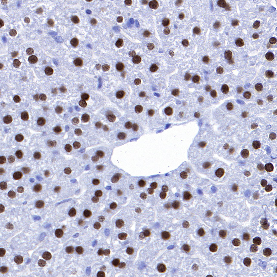
免疫组化
IHC shows positive staining in paraffin-embedded human stomach. Anti-FOXA2 antibody was used at 1/500 dilution, followed by a HRP Polymer for Mouse & Rabbit IgG (ready to use). Counterstained with hematoxylin. Heat mediated antigen retrieval with Tris/EDTA buffer pH9.0 was performed before commencing with IHC staining protocol.
(Weak affinity compared to mice)IHC shows positive staining in paraffin-embedded mouse liver. Anti-FOXA2 antibody was used at 1/500 dilution, followed by a HRP Polymer for Mouse & Rabbit IgG (ready to use). Counterstained with hematoxylin. Heat mediated antigen retrieval with Tris/EDTA buffer pH9.0 was performed before commencing with IHC staining protocol.
IHC shows positive staining in paraffin-embedded mouse lung. Anti-FOXA2 antibody was used at 1/500 dilution, followed by a HRP Polymer for Mouse & Rabbit IgG (ready to use). Counterstained with hematoxylin. Heat mediated antigen retrieval with Tris/EDTA buffer pH9.0 was performed before commencing with IHC staining protocol.
IHC shows positive staining in paraffin-embedded mouse stomach. Anti-FOXA2 antibody was used at 1/500 dilution, followed by a HRP Polymer for Mouse & Rabbit IgG (ready to use). Counterstained with hematoxylin. Heat mediated antigen retrieval with Tris/EDTA buffer pH9.0 was performed before commencing with IHC staining protocol.
IHC shows positive staining in paraffin-embedded rat liver. Anti-FOXA2 antibody was used at 1/200 dilution, followed by a HRP Polymer for Mouse & Rabbit IgG (ready to use). Counterstained with hematoxylin. Heat mediated antigen retrieval with Tris/EDTA buffer pH9.0 was performed before commencing with IHC staining protocol.
IHC shows positive staining in paraffin-embedded rat lung. Anti-FOXA2 antibody was used at 1/500 dilution, followed by a HRP Polymer for Mouse & Rabbit IgG (ready to use). Counterstained with hematoxylin. Heat mediated antigen retrieval with Tris/EDTA buffer pH9.0 was performed before commencing with IHC staining protocol.
IHC shows positive staining in paraffin-embedded rat stomach. Anti-FOXA2 antibody was used at 1/500 dilution, followed by a HRP Polymer for Mouse & Rabbit IgG (ready to use). Counterstained with hematoxylin. Heat mediated antigen retrieval with Tris/EDTA buffer pH9.0 was performed before commencing with IHC staining protocol.
免疫细胞化学
ICC shows positive staining in HepG2 cells. Anti-FOXA2 antibody was used at 1/500 dilution and incubated overnight at 4°C. Goat polyclonal Antibody to Rabbit IgG - H&L (Alexa Fluor® 488) was used as secondary antibody at 1/1000 dilution.The cells were fixed with 4% FPA and permeabilized with 0.1% PBS-Triton X-100. Nuclei were counterstained with DAPI.
Negative control: ICC shows negative staining in HeLa cells. Anti-FOXA2 antibody was used at 1/500 dilution and incubated overnight at 4°C. Goat polyclonal Antibody to Rabbit IgG - H&L (Alexa Fluor® 488) was used as secondary antibody at 1/1000 dilution.The cells were fixed with 4% FPA and permeabilized with 0.1% PBS-Triton X-100. Nuclei were counterstained with DAPI.
FAQs
我们一般不推荐客户回收利用抗体。 因为抗体使用之后缓冲体系已经发生改变,不同客户在回收抗体的保存条件上也会有差异,所以抗体回收使用效果无法保证。另外,我们对一批抗体回收验证测试,测试结果显示不同抗体可回收次数不同,一般效价越高的抗体,可重复使用的次数越多,客户可根据实验情况来确定

评论(0)