


 大包装询价
大包装询价 产品介绍 评论(0)
宿主来源
Rabbit抗原名称
CXCR5分子别名
C-X-C chemokine receptor type 5, BLR1, MDR15, CD185免疫原
Synthetic Peptide细胞定位
Cell membraneAccession
P32302克隆号
SDT-351-24抗体类型
Recombinant mAb应用
IHC-P, FCM, ICC反应种属 ?
Hu浓度
1 mg/ml性状
Liquid缓冲体系
PBS储存条件
12 months from date of receipt / reconstitution, 4°C as supplied
| 应用 | 稀释度 |
|---|---|
| IHC-P | 1:5000 |
| ICC | 1:100 |
| FCM | 1:500 |
CXCR5 is the receptor for the chemokine CXCL13, the chemokine important for secondary lymphoid tissue orchestration and lymphoid neogenesis. CXCL13 is constitutively expressed in secondary lymphoid tissue primarily by follicular dendritic cells in the spleen, lymph nodes, tonsils, and Peyer's patches, whereas CXCR5 is highly expressed in mature B lymphocytes and a subpopulation of follicular B helper T cells (Tfh). CXCL13 and its receptor CXCR5 are essential for trafficking B cells and homing B cells to lymphoid tissues and for the embryonic development of the majority of lymph nodes and Peyer's patches.
流式分析
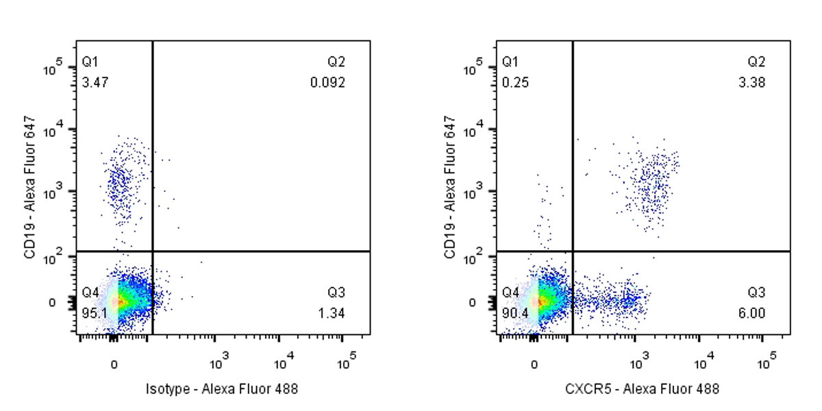
Flow cytometric analysis of Human peripheral blood monocytes labeling CXCR5 antibody at 1/500 dilution (0.1 μg) (Right) compared with a Rabbit monoclonal IgG isotype control (Left). Goat anti-Rabbit IgG(H+L) (Alexa Fluor® 488 Conjugate) was used as the secondary antibody at 1/2000 dilution.
Cells were surface stained with CD19-Alexa Fluor® 647, then stained with rabbit IgG (Left) / anti-CXCR5 (Right) separately. Gated on total viable cells.
免疫组化
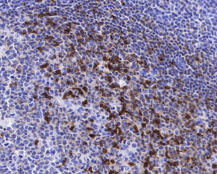
IHC shows positive staining in paraffin-embedded human tonsil. Anti-CXCR5 antibody was used at 1/5000 dilution, followed by a HRP Polymer for Mouse & Rabbit IgG (ready to use). Counterstained with hematoxylin. Heat mediated antigen retrieval with Tris/EDTA buffer pH9.0 was performed before commencing with IHC staining protocol.
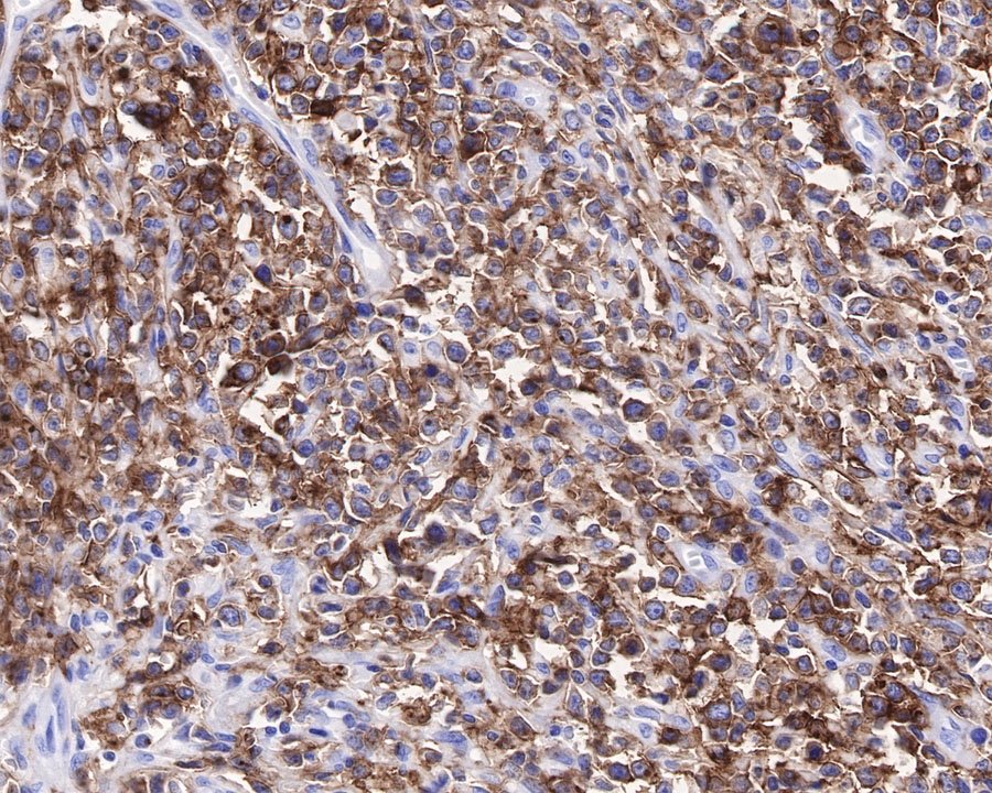
IHC shows positive staining in paraffin-embedded human diffuse large B-cell lymphoma. Anti-CXCR5 antibody was used at 1/5000 dilution, followed by a HRP Polymer for Mouse & Rabbit IgG (ready to use). Counterstained with hematoxylin. Heat mediated antigen retrieval with Tris/EDTA buffer pH9.0 was performed before commencing with IHC staining protocol.
免疫细胞化学
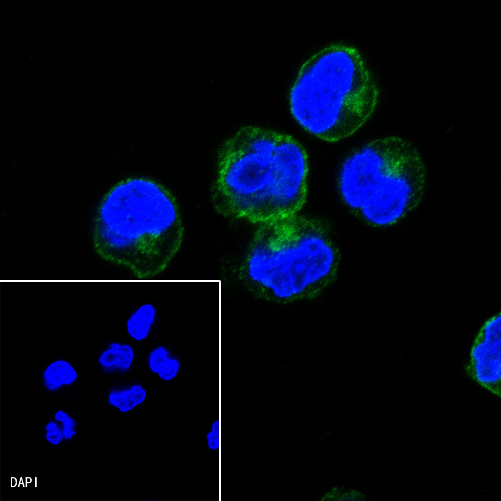
ICC shows positive staining in Raji cells. Anti-CXCR5 antibody was used at 1/100 dilution (Green) and incubated overnight at 4°C. Goat polyclonal Antibody to Rabbit IgG - H&L (Alexa Fluor® 488) was used as secondary antibody at 1/1000 dilution. The cells were fixed with 4% PFA and permeabilized with 0.1% PBS-Triton X-100. Nuclei were counterstained with DAPI (Blue).

Negative control: ICC shows negative staining in jurkat cells. Anti-CXCR5 antibody was used at 1/100 dilution (Green) and incubated overnight at 4°C. Goat polyclonal Antibody to Rabbit IgG - H&L (Alexa Fluor® 488) was used as secondary antibody at 1/1000 dilution. The cells were fixed with 4% PFA and permeabilized with 0.1% PBS-Triton X-100. Nuclei were counterstained with DAPI (Blue).

评论(0)