


 大包装询价
大包装询价 产品介绍 评论(0)
宿主来源
Rabbit抗原名称
HDAC1分子别名
Histone deacetylase 1, HD1, Protein deacetylase HDAC1免疫原
Synthetic Peptide细胞定位
NucleusAccession
Q13547克隆号
SDT-310-72抗体类型
Recombinant mAb应用
ICFCM, IHC-P, ICC, WB, IP反应种属 ?
Hu, Ms, Rt预测反应种属
(反应种属缩写表)Bv纯化方式
Protein A浓度
0.05 mg/ml性状
Liquid缓冲体系
PBS, 40% Glycerol, 0.05% BSA, 0.03% Proclin 300
储存条件
12 months from date of receipt / reconstitution, -20 °C as supplied.
| 应用 | 稀释度 |
|---|---|
| WB | 1:1000-1:5000 |
| IHC-P | 1:200 |
| ICC | 1:50 |
| ICFCM | 1:50 |
| IP | 1:25 |
HDAC family has four subclasses including I, II, III and IV. Histone deacetylase 1 (HDAC1) is a unique member of the classes I HDACs that has been shown to be involved in gene transcription, transcriptional regulation, cell cycle progression and developmental events by controlling both enzyme activity and epigenetics of key proteins [PMID: 16289629]. HDAC1 is the most abundant member of the class I HDACs in pulmonary endothelial cells [PMID: 34264338], regulating the enzymatic activity and epigenetics of key proteins to adapt to external stimuli. It can efficiently decrotonylate this relatively less abundant histone modification [PMID: 29317660].
免疫印迹
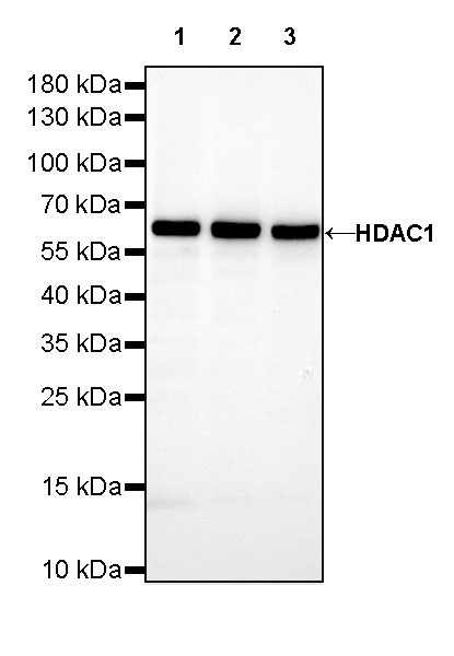 WB result of HDAC1 Rabbit mAb Primary antibody: HDAC1 Rabbit mAb at 1/5000 dilution Lane 1: HeLa whole cell lysate 20 µg Lane 2: Jurkat whole cell lysate 20 µg Lane 3: K-562 whole cell lysate 20 µg Secondary antibody: Goat Anti-Rabbit IgG, (H+L), HRP conjugated at 1/10000 dilution Predicted MW: 55kDa Observed MW: 62kDa
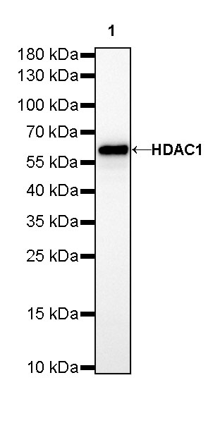 WB result of HDAC1 Rabbit mAb Primary antibody: HDAC1 Rabbit mAb at 1/5000 dilution Lane 1: NIH/3T3 whole cell lysate 20 µg Secondary antibody: Goat Anti-Rabbit IgG, (H+L), HRP conjugated at 1/10000 dilution Predicted MW: 55kDa Observed MW: 62kDa
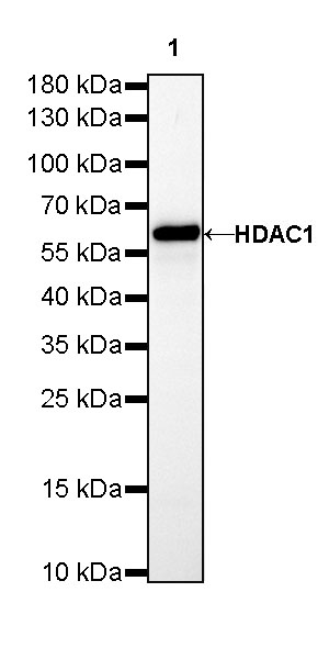 WB result of HDAC1 Rabbit mAb Primary antibody: HDAC1 Rabbit mAb at 1/5000 dilution Lane 1: C6 whole cell lysate 20 µg Secondary antibody: Goat Anti-Rabbit IgG, (H+L), HRP conjugated at 1/10000 dilution Predicted MW: 55kDa Observed MW: 62kDa
流式分析
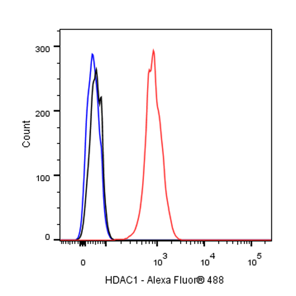
Flow cytometric analysis of 4% PFA fixed 90% methanol permeabilized HeLa (Human cervix adenocarcinoma epithelial cell) cells labelling HDAC1 antibody at 1/50 dilution (0.1 μg)/ (Red) compared with a Rabbit monoclonal IgG (Black) isotype control and an unlabelled control (cells without incubation with primary antibody and secondary antibody) (Blue). Goat Anti - Rabbit IgG Alexa Fluor® 488 was used as the secondary antibody.
免疫沉淀
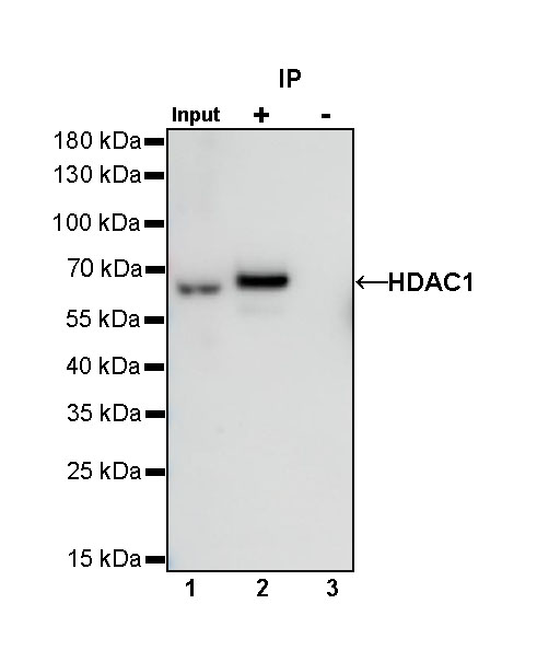
HDAC1 Rabbit mAb at 1/25 dilution (0.2 µg) immunoprecipitating HDAC1 in 0.2 mg HeLa whole cell lysate.
Western blot was performed on the immunoprecipitate using HDAC1 Rabbit mAb at 1/1000 dilution.
Secondary antibody (HRP) for IP was used at 1/400 dilution.
Lane 1: HeLa whole cell lysate 5 µg (Input)
Lane 2: HDAC1 Rabbit mAb IP in HeLa whole cell lysate
Lane 3: Rabbit monoclonal IgG IP in HeLa whole cell lysate
Predicted MW: 55 kDa
Observed MW: 62 kDa
免疫组化
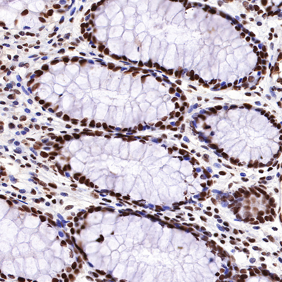 IHC shows positive staining in paraffin-embedded human colon. Anti-HDAC1 antibody was used at 1/200 dilution, followed by a HRP Polymer for Mouse & Rabbit IgG (ready to use). Counterstained with hematoxylin. Heat mediated antigen retrieval with Tris/EDTA buffer pH9.0 was performed before commencing with IHC staining protocol.
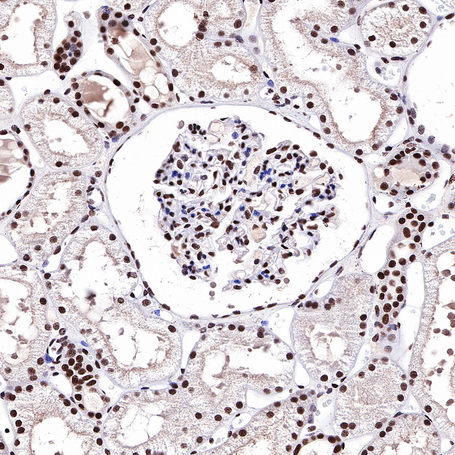 IHC shows positive staining in paraffin-embedded human kidney. Anti-HDAC1 antibody was used at 1/200 dilution, followed by a HRP Polymer for Mouse & Rabbit IgG (ready to use). Counterstained with hematoxylin. Heat mediated antigen retrieval with Tris/EDTA buffer pH9.0 was performed before commencing with IHC staining protocol.
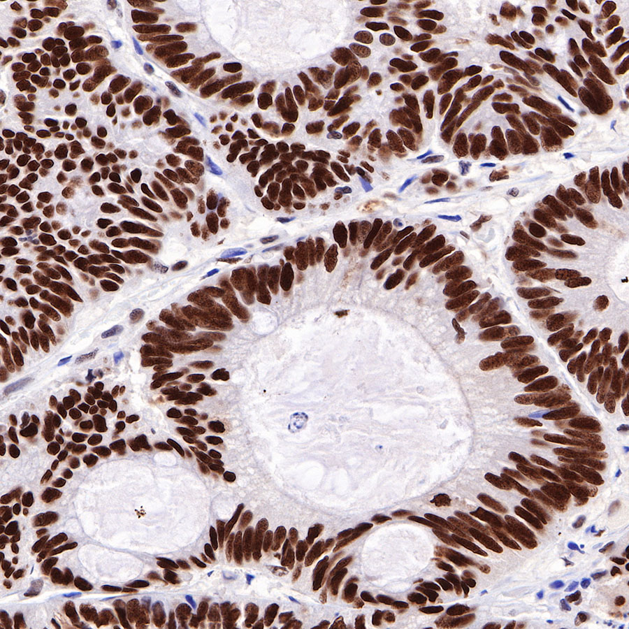 IHC shows positive staining in paraffin-embedded human colon cancer. Anti-HDAC1 antibody was used at 1/200 dilution, followed by a HRP Polymer for Mouse & Rabbit IgG (ready to use). Counterstained with hematoxylin. Heat mediated antigen retrieval with Tris/EDTA buffer pH9.0 was performed before commencing with IHC staining protocol.
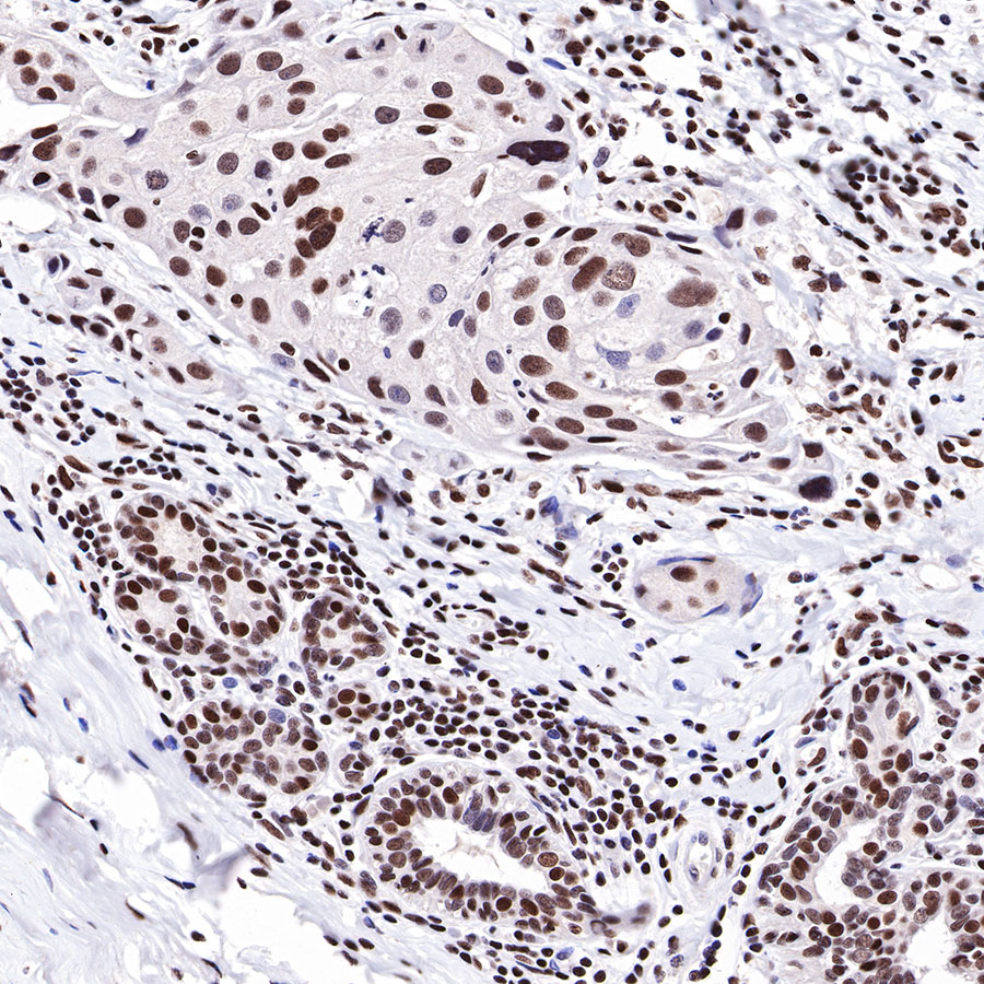 IHC shows positive staining in paraffin-embedded human breast cancer. Anti-HDAC1 antibody was used at 1/200 dilution, followed by a HRP Polymer for Mouse & Rabbit IgG (ready to use). Counterstained with hematoxylin. Heat mediated antigen retrieval with Tris/EDTA buffer pH9.0 was performed before commencing with IHC staining protocol.
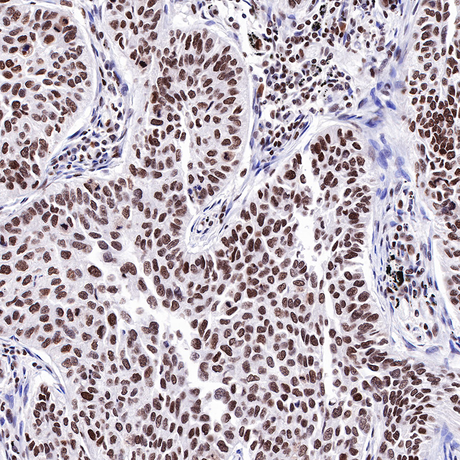 IHC shows positive staining in paraffin-embedded human lung squamous cell carcinoma. Anti-HDAC1 antibody was used at 1/200 dilution, followed by a HRP Polymer for Mouse & Rabbit IgG (ready to use). Counterstained with hematoxylin. Heat mediated antigen retrieval with Tris/EDTA buffer pH9.0 was performed before commencing with IHC staining protocol.
免疫细胞化学

ICC shows positive staining in HeLa cells. Anti-HDAC1 antibody was used at 1/50 dilution (Green) and incubated overnight at 4°C. Goat polyclonal Antibody to Rabbit IgG - H&L (Alexa Fluor® 488) was used as secondary antibody at 1/1000 dilution. The cells were fixed with 100% ice-cold methanol and permeabilized with 0.1% PBS-Triton X-100. Nuclei were counterstained with DAPI (Blue). Counterstain with tubulin (red).

评论(0)