


 大包装询价
大包装询价 产品介绍 评论(0)
宿主来源
Rabbit抗原名称
Beta III tubulin分子别名
Tubulin beta-3 chain, Tubulin beta-4 chain, Tubulin beta-III免疫原
Synthetic Peptide细胞定位
Cytoplasm, CytoskeletonAccession
Q13509克隆号
SDT-251-28抗体类型
Recombinant mAb应用
ICFCM, IHC-P, ICC, WB, IP反应种属 ?
Hu, Ms, Rt纯化方式
Protein A浓度
0.05 mg/ml性状
Liquid缓冲体系
PBS, 40% Glycerol, 0.05% BSA, 0.03% Proclin 300储存条件
12 months from date of receipt / reconstitution, -20 °C as supplied.
| 应用 | 稀释度 |
|---|---|
| WB | 1:1000-1:5000 |
| IHC-P | 1:200 |
| ICFCM | 1:50 |
| ICC | 1:50 |
| IP | 1:25 |
Beta III-tubulin is frequently overexpressed in human tumors and is associated with resistance to microtubule-targeting agents, tumor aggressiveness, and poor patient outcome [PMID: 34364992IF: 9.7 Q1 ]. In prostate cancer, βIII-tubulin expression is strongly associated with both TMPRSS2: ERG rearrangement, ERG expression and PTEN deletions, three key oncogenetic features of aggressive prostate cancer [PubMed: 24378408]. Also, colorectal cancer is more aggressive in young male patients in whom testosterone elevation and activation of TUBB3 are connected with poor outcome [PubMed: 23637935].
免疫印迹
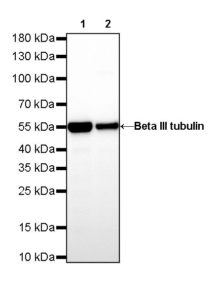 WB result of Beta III tubulin Rabbit mAb Primary antibody: Beta III tubulin Rabbit mAb at 1/5000 dilution Lane 1: SH-SY5Y whole cell lysate 20 µg Lane 2: U-87 MG whole cell lysate 20 µg Secondary antibody: Goat Anti-Rabbit IgG, (H+L), HRP conjugated at 1/10000 dilution Predicted MW: 50kDa Observed MW: 55kDa
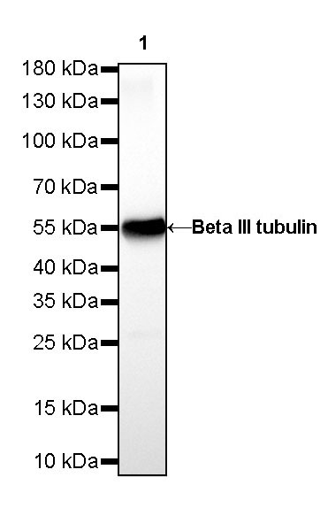 WB result of Beta III tubulin Rabbit mAb Primary antibody: Beta III tubulin Rabbit mAb at 1/5000 dilution Lane 1: mouse brain lysate 20 µg Secondary antibody: Goat Anti-Rabbit IgG, (H+L), HRP conjugated at 1/10000 dilution Predicted MW: 50kDa Observed MW: 55kDa
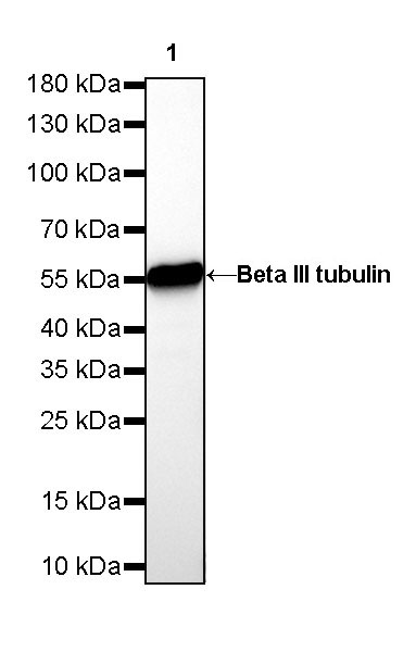 WB result of Beta III tubulin Rabbit mAb Primary antibody: Beta III tubulin Rabbit mAb at 1/5000 dilution Lane 1: rat brain lysate 20 µg Secondary antibody: Goat Anti-Rabbit IgG, (H+L), HRP conjugated at 1/10000 dilution Predicted MW: 50kDa Observed MW: 55kDa
流式分析
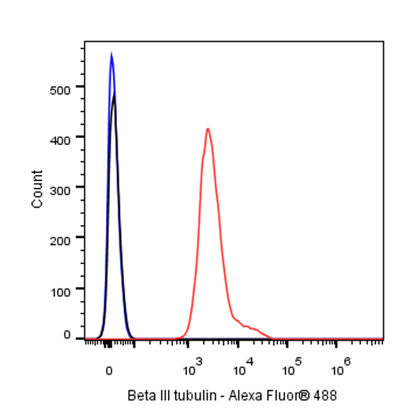
Flow cytometric analysis of 4% PFA fixed 90% methanol permeabilized SH-SY5Y (Human neuroblastoma epithelial cell) cells labelling Beta III tubulin antibody at 1/50 (0.1 μg) dilution / (red) compared with a Rabbit monoclonal IgG (Black) isotype control and an unlabelled control (cells without incubation with primary antibody and secondary antibody) (Blue). Goat Anti - Rabbit IgG Alexa Fluor® 488 was used as the secondary antibody.
免疫沉淀
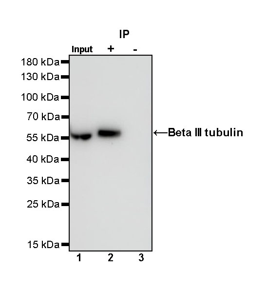
Beta III tubulin Rabbit mAb at 1/25 dilution (0.2 µg) immunoprecipitating Beta III tubulin in 0.2 mg SH-SY5Y whole cell lysate.
Western blot was performed on the immunoprecipitate using Beta III tubulin Rabbit mAb at 1/1000 dilution.
Secondary antibody (HRP) for IP was used at 1/400 dilution.
Lane 1: SH-SY5Y whole cell lysate 5 µg (Input)
Lane 2: Beta III tubulin Rabbit mAb IP in SH-SY5Y whole cell lysate
Lane 3: Rabbit monoclonal IgG IP in SH-SY5Y whole cell lysate
Predicted MW: 50 kDa
Observed MW: 55 kDa
免疫组化
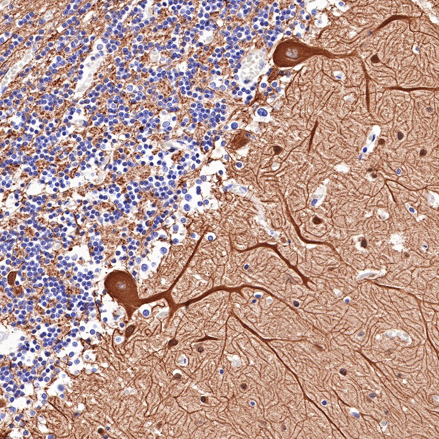 IHC shows positive staining in paraffin-embedded human cerebellum. Anti-Beta III tubulin antibody was used at 1/200 dilution, followed by a HRP Polymer for Mouse & Rabbit IgG (ready to use). Counterstained with hematoxylin. Heat mediated antigen retrieval with Tris/EDTA buffer pH9.0 was performed before commencing with IHC staining protocol.
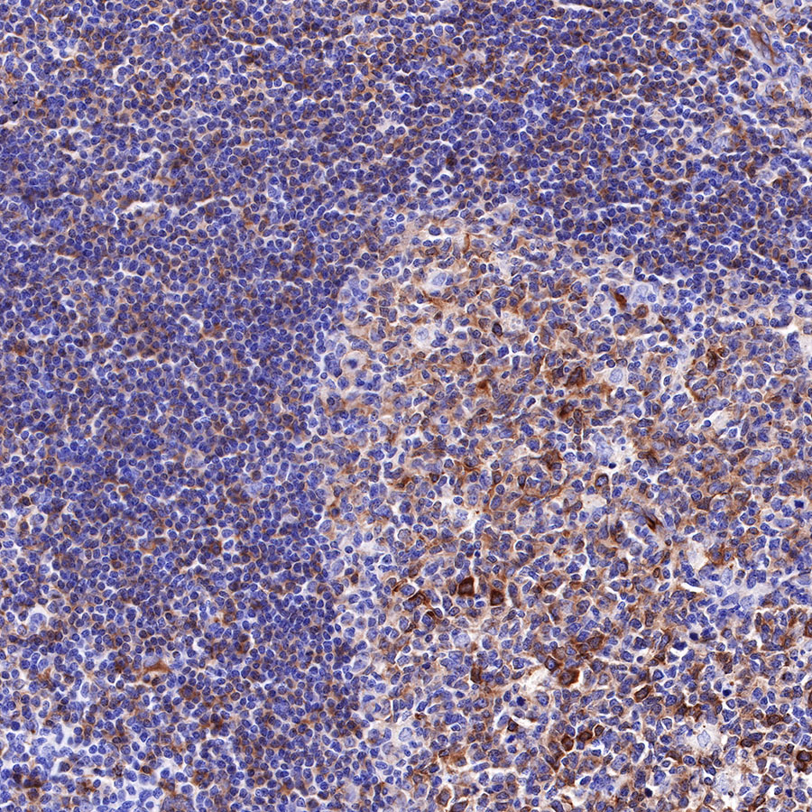 IHC shows positive staining in paraffin-embedded human tonsil. Anti-Beta III tubulin antibody was used at 1/200 dilution, followed by a HRP Polymer for Mouse & Rabbit IgG (ready to use). Counterstained with hematoxylin. Heat mediated antigen retrieval with Tris/EDTA buffer pH9.0 was performed before commencing with IHC staining protocol.
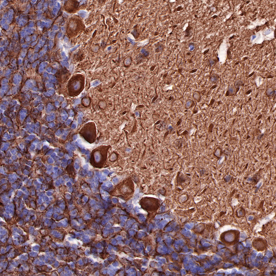 IHC shows positive staining in paraffin-embedded mouse cerebellum. Anti-Beta III tubulin antibody was used at 1/200 dilution, followed by a HRP Polymer for Mouse & Rabbit IgG (ready to use). Counterstained with hematoxylin. Heat mediated antigen retrieval with Tris/EDTA buffer pH9.0 was performed before commencing with IHC staining protocol.
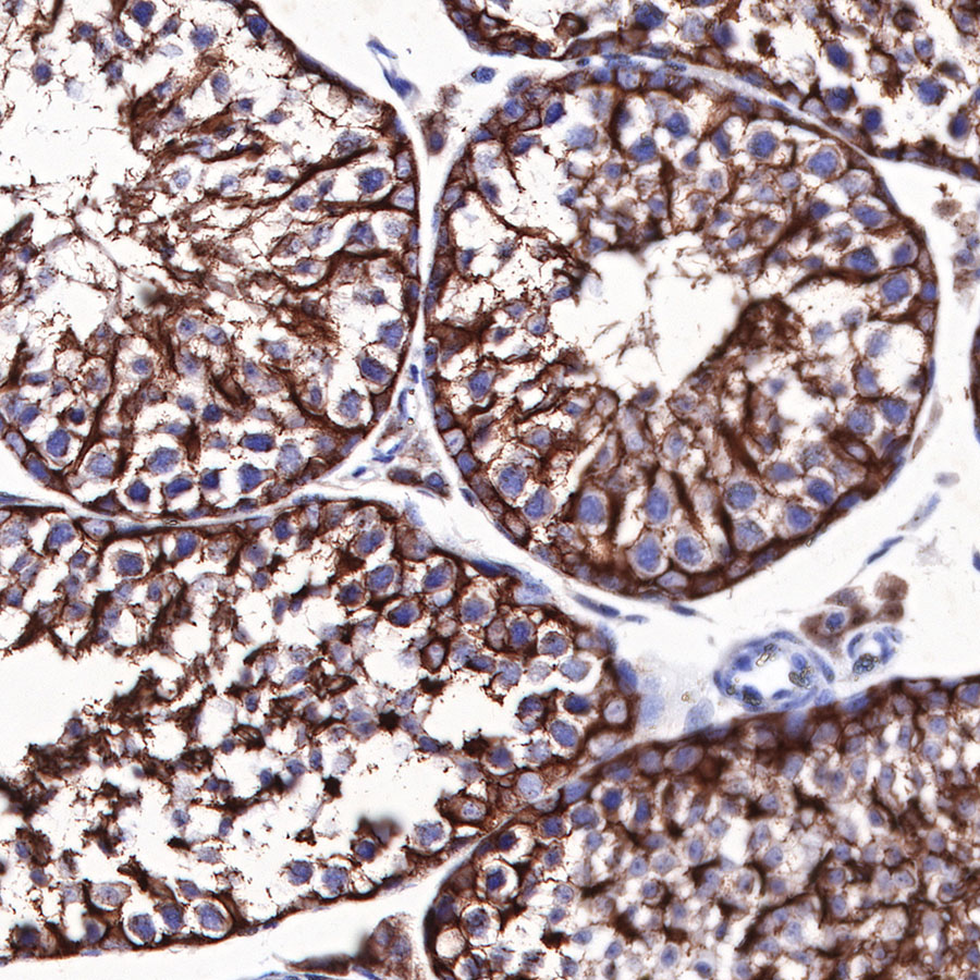 IHC shows positive staining in paraffin-embedded mouse testis. Anti-Beta III tubulin antibody was used at 1/200 dilution, followed by a HRP Polymer for Mouse & Rabbit IgG (ready to use). Counterstained with hematoxylin. Heat mediated antigen retrieval with Tris/EDTA buffer pH9.0 was performed before commencing with IHC staining protocol.
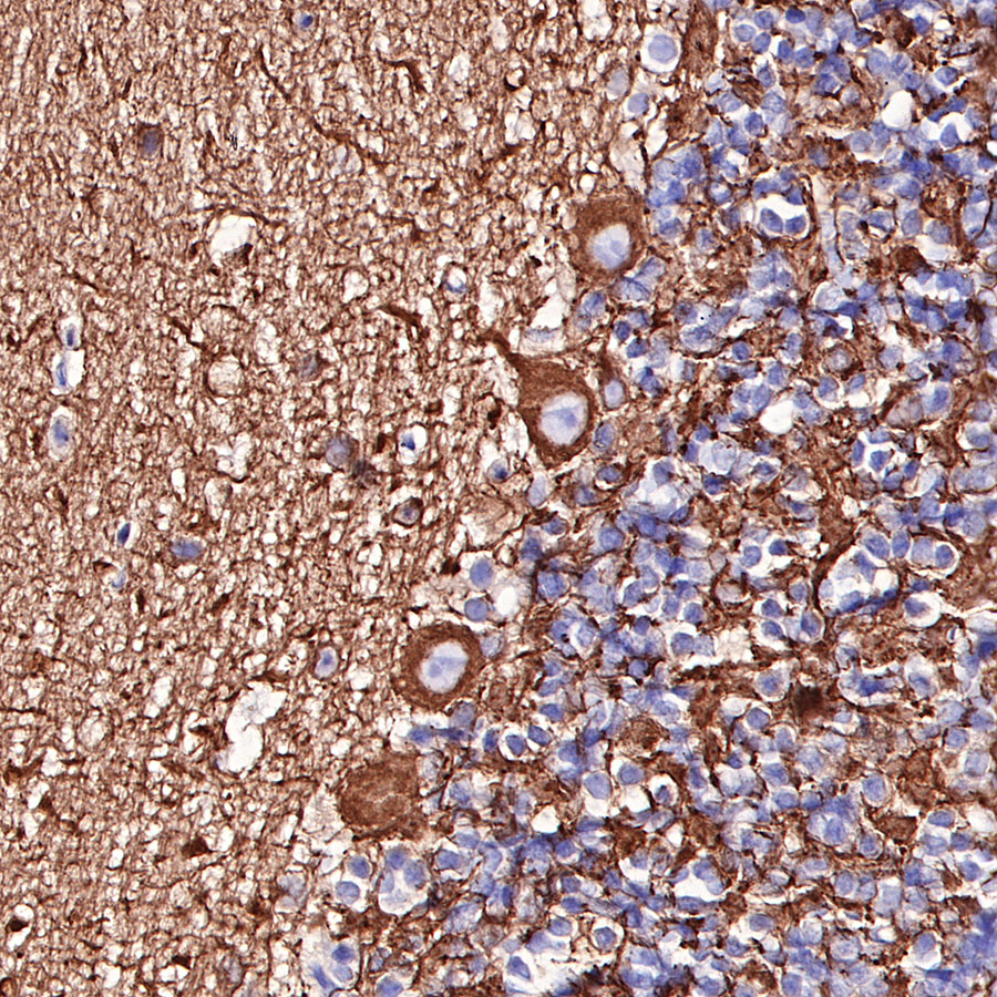 IHC shows positive staining in paraffin-embedded rat cerebellum. Anti-Beta III tubulin antibody was used at 1/200 dilution, followed by a HRP Polymer for Mouse & Rabbit IgG (ready to use). Counterstained with hematoxylin. Heat mediated antigen retrieval with Tris/EDTA buffer pH9.0 was performed before commencing with IHC staining protocol.
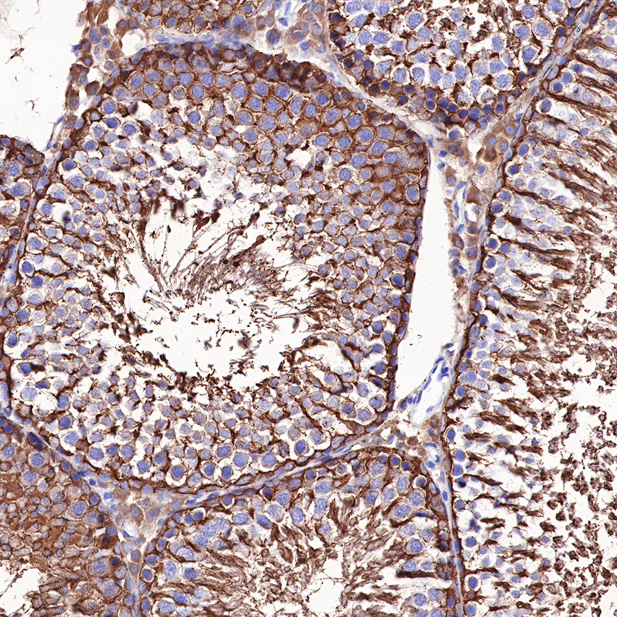 IHC shows positive staining in paraffin-embedded rat testis. Anti-Beta III tubulin antibody was used at 1/200 dilution, followed by a HRP Polymer for Mouse & Rabbit IgG (ready to use). Counterstained with hematoxylin. Heat mediated antigen retrieval with Tris/EDTA buffer pH9.0 was performed before commencing with IHC staining protocol.
免疫细胞化学
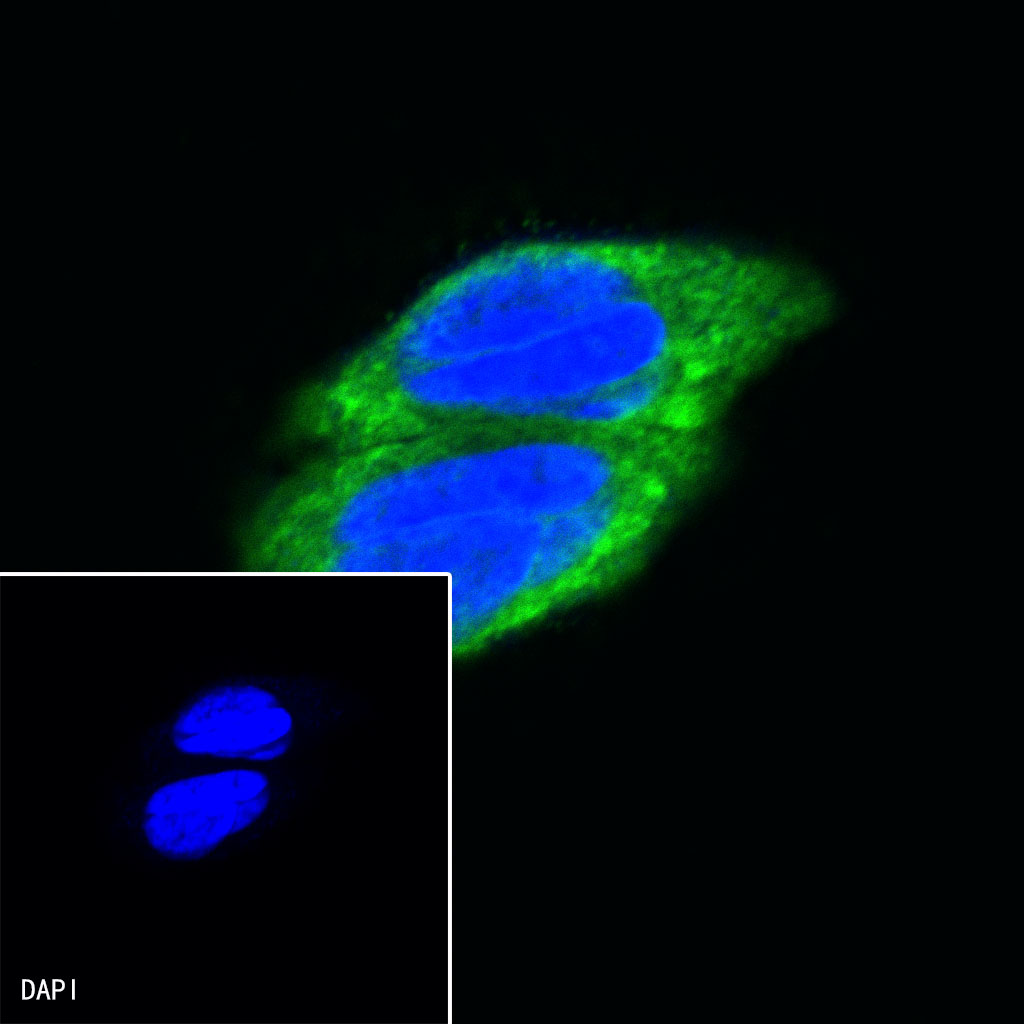
ICC shows positive staining in SH-SY5Y cells. Anti-Beta III tubulin antibody was used at 1/50 dilution (Green) and incubated overnight at 4°C. Goat polyclonal Antibody to Rabbit IgG - H&L (Alexa Fluor® 488) was used as secondary antibody at 1/1000 dilution. The cells were fixed with 100% ice-cold methanol and permeabilized with 0.1% PBS-Triton X-100. Nuclei were counterstained with DAPI (Blue).

评论(0)