


 大包装询价
大包装询价 产品介绍 评论(0)
宿主来源
Rabbit抗原名称
Ubiquitin分子别名
N/A免疫原
N/A细胞定位
Cytoplasm, NucleusAccession
P62987克隆号
SDT-R095抗体类型
Recombinant mAb应用
ICFCM, IHC-P, ICC, WB反应种属 ?
Species Independent纯化方式
Protein A浓度
0.5 mg/ml性状
Liquid缓冲体系
PBS, 40% Glycerol, 0.05% BSA, 0.03% Proclin 300
储存条件
12 months from date of receipt / reconstitution, -20 °C as supplied
| 应用 | 稀释度 |
|---|---|
| WB | 1:1000 |
| IHC-P | 1:2000 |
| ICFCM | 1:500 |
| ICC | 1:500 |
Protein ubiquitination is a dynamic multifaceted post-translational modification involved in nearly all aspects of eukaryotic biology. Once attached to a substrate, the 76-amino acid protein ubiquitin is subjected to further modifications, creating a multitude of distinct signals with distinct cellular outcomes, referred to as the 'ubiquitin code'. Ubiquitin can be ubiquitinated on seven lysine (Lys) residues or on the N-terminus, leading to polyubiquitin chains that can encompass complex topologies. Alternatively or in addition, ubiquitin Lys residues can be modified by ubiquitin-like molecules (such as SUMO or NEDD8). Finally, ubiquitin can also be acetylated on Lys, or phosphorylated on Ser, Thr or Tyr residues, and each modification has the potential to dramatically alter the signaling outcome [PMID: 27012465].
免疫印迹

WB result of Ubiquitin Rabbit mAb
Primary antibody: Ubiquitin Rabbit mAb at 1/1000 dilution
Lane 1: untreated HT-1080 whole cell lysate 20 µg
Lane 2: HT-1080 treated with MG-132(10μM, 4h) whole cell lysate 20 µg
Secondary antibody: Goat Anti-Rabbit IgG, (H+L), HRP conjugated at 1/10000 dilution
Observed MW: 16~300 kDa
WB result of Ubiquitin Rabbit mAb
Primary antibody: Ubiquitin Rabbit mAb at 1/1000 dilution
Lane 1: untreated MCF7 whole cell lysate 20 µg
Lane 2: MCF7 treated with MG-132(10μM, 2h) whole cell lysate 20 µg
Secondary antibody: Goat Anti-Rabbit IgG, (H+L), HRP conjugated at 1/10000 dilution
Observed MW: 16~300 kDa
WB result of Ubiquitin Rabbit mAb
Primary antibody: Ubiquitin Rabbit mAb at 1/1000 dilution
Lane 1: NIH/3T3 whole cell lysate 20 µg
Secondary antibody: Goat Anti-Rabbit IgG, (H+L), HRP conjugated at 1/10000 dilution
Observed MW: 16~300 kDa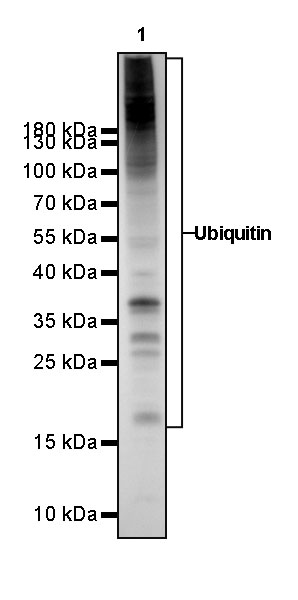
WB result of Ubiquitin Rabbit mAb
Primary antibody: Ubiquitin Rabbit mAb at 1/1000 dilution
Lane 1: C6 whole cell lysate 20 µg
Secondary antibody: Goat Anti-Rabbit IgG, (H+L), HRP conjugated at 1/10000 dilution
Observed MW: 16~300 kDa
流式分析
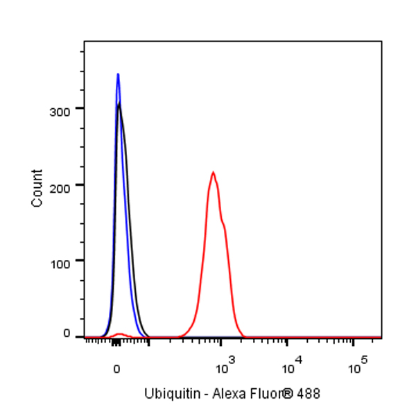
Flow cytometric analysis of 4% PFA fixed 90% methanol permeabilized C6 (Rat glial tumor glial cell) cells labelling Ubiquitin antibody at 1/500 dilution (0.1 μg) / (red) compared with a Rabbit monoclonal IgG (Black) isotype control and an unlabelled control (cells without incubation with primary antibody and secondary antibody) (Blue). Goat Anti - Rabbit IgG Alexa Fluor® 488 was used as the secondary antibody.
免疫组化
 IHC shows positive staining in paraffin-embedded human kidney. Anti-Ubiquitin antibody was used at 1/2000 dilution, followed by a HRP Polymer for Mouse & Rabbit IgG (ready to use). Counterstained with hematoxylin. Heat mediated antigen retrieval with Tris/EDTA buffer pH9.0 was performed before commencing with IHC staining protocol.
 IHC shows positive staining in paraffin-embedded human lung squamous cell carcinoma. Anti-Ubiquitin antibody was used at 1/2000 dilution, followed by a HRP Polymer for Mouse & Rabbit IgG (ready to use). Counterstained with hematoxylin. Heat mediated antigen retrieval with Tris/EDTA buffer pH9.0 was performed before commencing with IHC staining protocol.
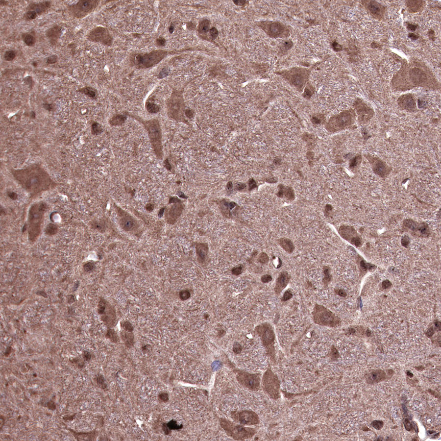 IHC shows positive staining in paraffin-embedded mouse cerebral cortex. Anti-Ubiquitin antibody was used at 1/2000 dilution, followed by a HRP Polymer for Mouse & Rabbit IgG (ready to use). Counterstained with hematoxylin. Heat mediated antigen retrieval with Tris/EDTA buffer pH9.0 was performed before commencing with IHC staining protocol.
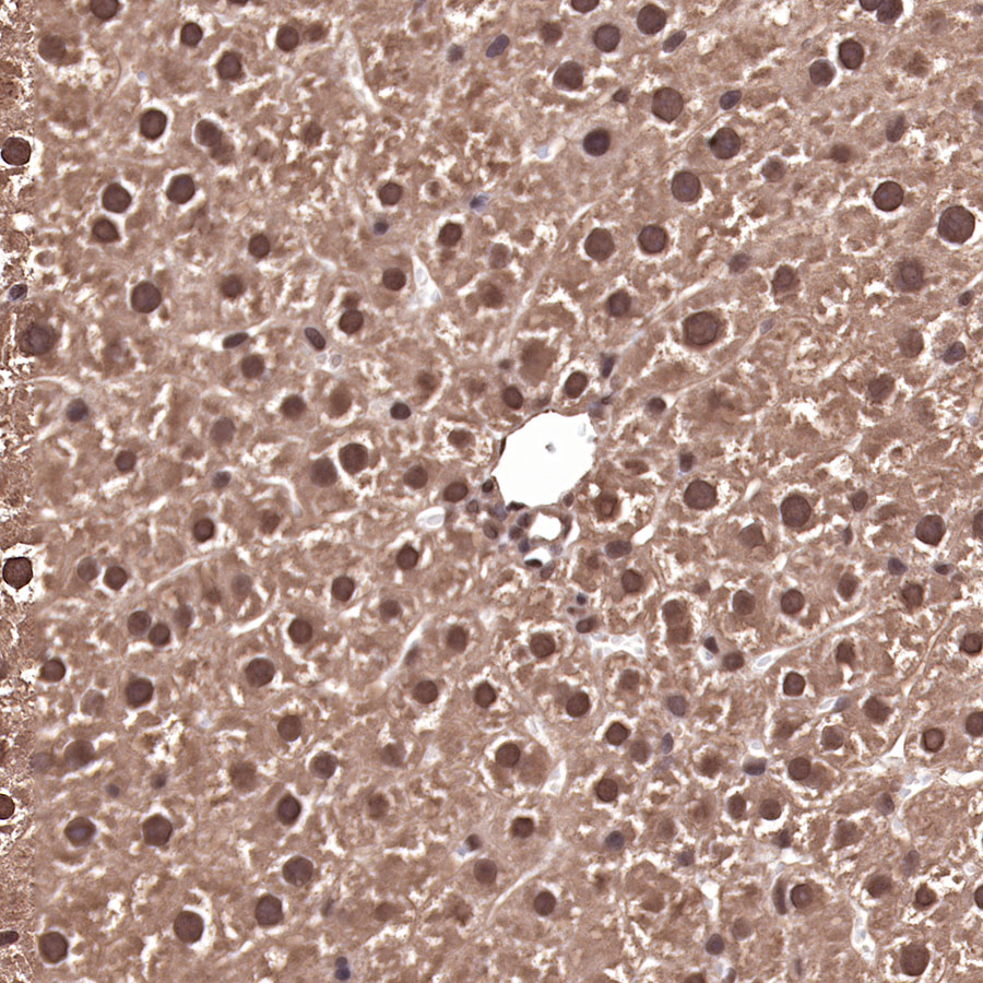 IHC shows positive staining in paraffin-embedded rat liver. Anti-Ubiquitin antibody was used at 1/2000 dilution, followed by a HRP Polymer for Mouse & Rabbit IgG (ready to use). Counterstained with hematoxylin. Heat mediated antigen retrieval with Tris/EDTA buffer pH9.0 was performed before commencing with IHC staining protocol.
免疫细胞化学
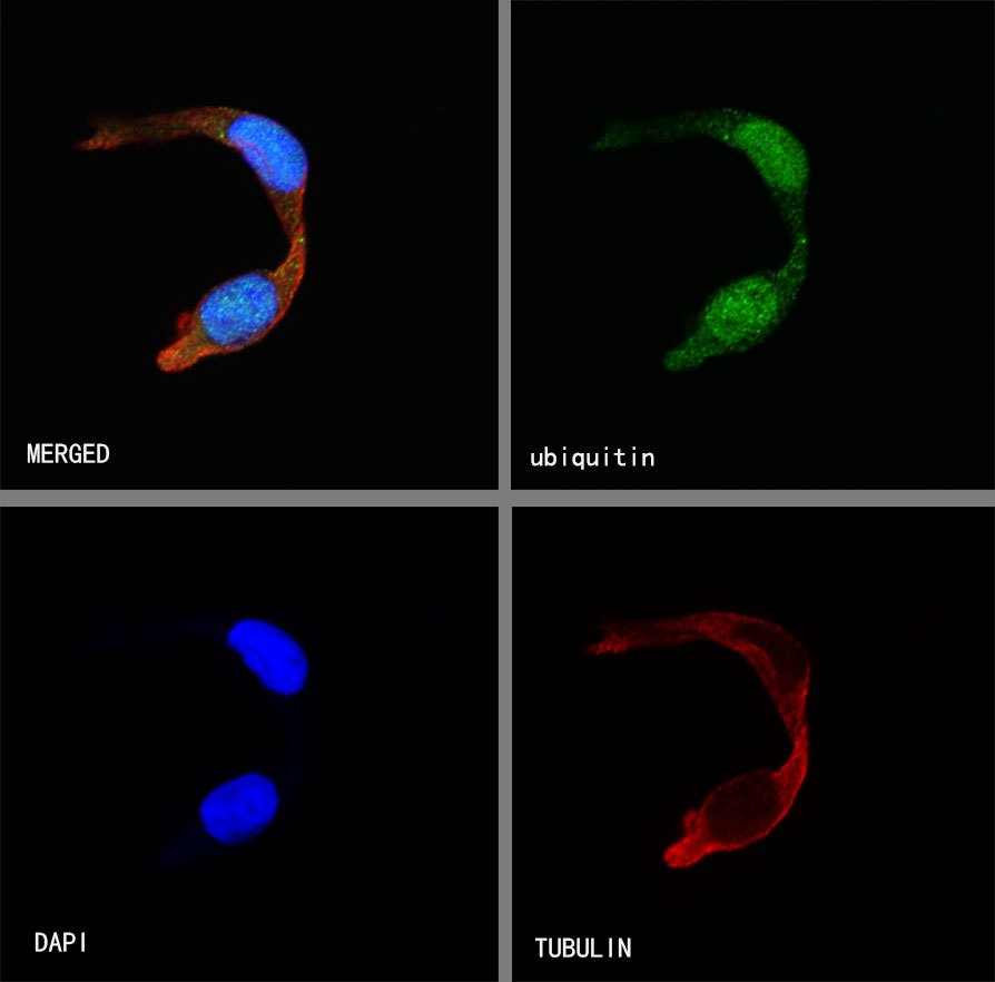
ICC shows positive staining in C6 cells. Anti-Ubiquitin antibody was used at 1/500 dilution (Green) and incubated overnight at 4°C. Goat polyclonal Antibody to Rabbit IgG - H&L (Alexa Fluor® 488) was used as secondary antibody at 1/1000 dilution. The cells were fixed with 100% ice-cold methanol and permeabilized with 0.1% PBS-Triton X-100. Nuclei were counterstained with DAPI (Blue). Counterstain with tubulin (red).

评论(0)