


产品介绍 引用文献(1) 评论(0)
宿主来源
Rabbit抗原名称
β-actin分子别名
ACTB免疫原
N/A细胞定位
Cytoplasm, CytoskeletonAccession
P60709克隆号
SDT-R156抗体类型
Recombinant mAb应用
ICFCM ,IHC-P ? ,ICC ? ,WB反应种属 ?
Hu, Ms纯化方式
Protein A浓度
0.5 mg/ml性状
Liquid缓冲体系
PBS, 40% Glycerol, 0.05% BSA, 0.03% Proclin 300
储存条件
12 months from date of receipt / reconstitution, -20 °C as supplied.
RRID
AB_3712962
| 应用 | 稀释度 |
|---|---|
| WB | 1:1000-1:5000 |
| IHC-P | 1:2000 |
| ICC | 1:500 |
| ICFCM | 1:500 |
Beta-actin (human gene and protein abbreviation ACTB/ACTB) is one of six different actin isoforms which have been identified in humans. This is one of the two non-muscle cytoskeletal actins. Actins are highly conserved proteins that are involved in cell motility, structure and integrity. Beta actin is often used in Western blotting as a loading control, to normalize total protein amounts and check for eventual protein degradation in the samples. Its transcript is also commonly used as a housekeeping gene standard in qPCR. Its molecular weight is approximately 42 kDa.
免疫印迹
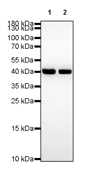
- WB result of β-actin Rabbit mAb Primary antibody: β-actin Rabbit mAb at 1/1000 dilution Lane 1: HeLa whole cell lysate 20 µg Lane 2: A549 whole cell lysate 20 µg Secondary antibody: Goat Anti-Rabbit IgG, (H+L), HRP conjugated at 1/10000 dilution Predicted MW: 42kDa Observed MW: 42kDa
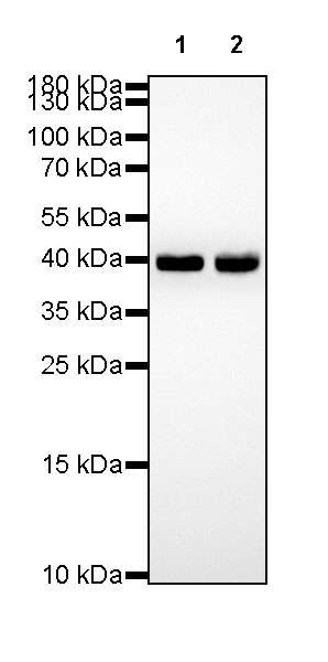
- WB result of β-actin Rabbit mAb Primary antibody: β-actin Rabbit mAb at 1/1000 dilution Lane 1: NIH/3T3 whole cell lysate 20 µg Lane 2: mouse kidney lysate 20 µg Secondary antibody: Goat Anti-Rabbit IgG, (H+L), HRP conjugated at 1/10000 dilution Predicted MW: 42kDa Observed MW: 42kDa
流式分析
- Flow cytometric analysis of HeLa cells labelling β-actin antibody at 1/500 (0.1 μg) dilution/ (red) compared with a Rabbit monoclonal IgG (Black) isotype control and an unlabelled control (cells without incubation with primary antibody and secondary antibody) (Blue). Goat Anti-Rabbit IgG Alexa Fluor® 488 was used as the secondary antibody.
- Flow cytometric analysis of NIH/3T3 cells labelling β-actin antibody at 1/500 (0.1 μg) dilution/ (red) compared with a Rabbit monoclonal IgG (Black) isotype control and an unlabelled control (cells without incubation with primary antibody and secondary antibody) (Blue). Goat Anti-Rabbit IgG Alexa Fluor® 488 was used as the secondary antibody.
免疫组化
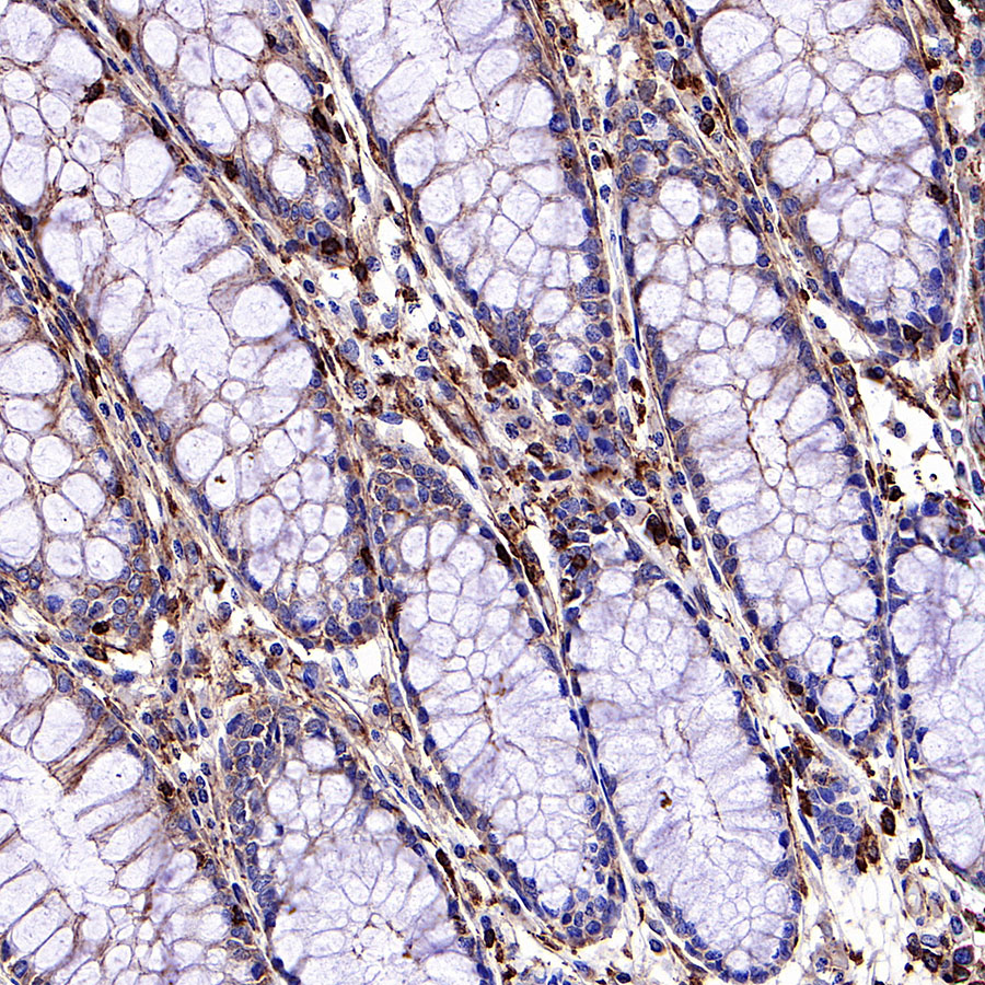
- IHC shows positive staining in paraffin-embedded human colon. Anti-β-actin antibody was used at 1/2000 dilution, followed by a HRP Polymer for Mouse & Rabbit IgG (ready to use). Counterstained with hematoxylin. Heat mediated antigen retrieval with Tris/EDTA buffer pH9.0 was performed before commencing with IHC staining protocol.
- IHC shows positive staining in paraffin-embedded human cardiac muscle. Anti-β-actin antibody was used at 1/2000 dilution, followed by a HRP Polymer for Mouse & Rabbit IgG (ready to use). Counterstained with hematoxylin. Heat mediated antigen retrieval with Tris/EDTA buffer pH9.0 was performed before commencing with IHC staining protocol.
- IHC shows positive staining in paraffin-embedded human colon cancer. Anti-β-actin antibody was used at 1/2000 dilution, followed by a HRP Polymer for Mouse & Rabbit IgG (ready to use). Counterstained with hematoxylin. Heat mediated antigen retrieval with Tris/EDTA buffer pH9.0 was performed before commencing with IHC staining protocol.
- IHC shows positive staining in paraffin-embedded human hepatocellular carcinoma. Anti-β-actin antibody was used at 1/2000 dilution, followed by a HRP Polymer for Mouse & Rabbit IgG (ready to use). Counterstained with hematoxylin. Heat mediated antigen retrieval with Tris/EDTA buffer pH9.0 was performed before commencing with IHC staining protocol.
- IHC shows positive staining in paraffin-embedded human lung squamous cell carcinoma. Anti-β-actin antibody was used at 1/2000 dilution, followed by a HRP Polymer for Mouse & Rabbit IgG (ready to use). Counterstained with hematoxylin. Heat mediated antigen retrieval with Tris/EDTA buffer pH9.0 was performed before commencing with IHC staining protocol.
- IHC shows positive staining in paraffin-embedded mouse kidney. Anti-β-actin antibody was used at 1/2000 dilution, followed by a HRP Polymer for Mouse & Rabbit IgG (ready to use). Counterstained with hematoxylin. Heat mediated antigen retrieval with Tris/EDTA buffer pH9.0 was performed before commencing with IHC staining protocol.
免疫细胞化学
- ICC shows positive staining in HeLa cells. Anti-β-actin antibody was used at 1/500 dilution (Green) and incubated overnight at 4°C. Goat polyclonal Antibody to Rabbit IgG - H&L (Alexa Fluor® 488) was used as secondary antibody at 1/1000 dilution. The cells were fixed with 100% ice-cold methanol and permeabilized with 0.1% PBS-Triton X-100. Nuclei were counterstained with DAPI (Blue). Counterstain with tubulin (red).
- ICC shows positive staining in NIH/3T3 cells. Anti-β-actin antibody was used at 1/500 dilution (Green) and incubated overnight at 4°C. Goat polyclonal Antibody to Rabbit IgG - H&L (Alexa Fluor® 488) was used as secondary antibody at 1/1000 dilution. The cells were fixed with 100% ice-cold methanol and permeabilized with 0.1% PBS-Triton X-100. Nuclei were counterstained with DAPI (Blue). Counterstain with tubulin (red).
引用文献(1)
- Cancer-associated adipocytes mediate CD8+ T cell dysfunction via FGF21-driven lipolysis
S Dalangood, C Hu, C Yuan, X Li, W Qiao, H Li
BioRxiv. 2024 Oct 21 .
货号:S0B0074产品名称:β-actin Recombinant Rabbit mAb (SDT-R156)

评论(0)