


产品介绍 评论(0)
宿主来源
Rabbit抗原名称
α-Actinin-1分子别名
Alpha-actinin-1, ACTN1免疫原
Synthetic Peptide细胞定位
Cytoskeleton, Cell membrane, CytoplasmAccession
P12814克隆号
SDT-311-20抗体类型
Rabbit mAb应用
IHC-P ? ,WB反应种属 ?
Hu, Ms, Rt预测反应种属
(反应种属缩写表)Bv, Mq纯化方式
Protein A浓度
0.5 mg/ml性状
Liquid缓冲体系
PBS, 40% Glycerol, 0.05% BSA, 0.03% Proclin 300
储存条件
12 months from date of receipt / reconstitution, -20 °C as supplied.
| 应用 | 稀释度 |
|---|---|
| WB | 1:1000-1:5000 |
| IHC-P | 1:2000 |
Alpha-actinin (α-actinin) is a ubiquitous cytoskeletal protein, which belongs to the superfamily of filamentous actin (F-actin) crosslinking proteins. It is present in multiple subcellular regions of both muscle and non-muscle cells, including cell-cell and cell-matrix contact sites, cellular protrusions and stress fiber dense regions and thus, it seems to bear multiple important roles in the cell by linking the cytoskeleton to many different transmembrane proteins in a variety of junctions. Four isoforms of human α-actinin have already been identified namely, the "muscles" α-actinin-2 and α-actinin-3 and the "non-muscles" α-actinin-1 and α-actinin-4 [PMID: 21241830]. α-actinin-1 is an important intracellular calcium-sensing protein, the major actin crosslinking protein in focal adhesions and stress fibers [PMID: 27272015].
免疫印迹
WB result of α-Actinin-1 Rabbit mAb
Primary antibody: α-Actinin-1 Rabbit mAb at 1/1000 dilution
Lane 1: HepG2 whole cell lysate 20 µg
Lane 2: HeLa whole cell lysate 20 µg
Secondary antibody: Goat Anti-Rabbit IgG, (H+L), HRP conjugated at 1/10000 dilution
Predicted MW: 103 kDa
Observed MW: 103 kDaWB result of α-Actinin-1 Rabbit mAb
Primary antibody: α-Actinin-1 Rabbit mAb at 1/1000 dilution
Lane 1: mouse liver lysate 20 µg
Lane 2: NIH/3T3 whole cell lysate 20 µg
Secondary antibody: Goat Anti-Rabbit IgG, (H+L), HRP conjugated at 1/10000 dilution
Predicted MW: 103 kDa
Observed MW: 103 kDaWB result of α-Actinin-1 Rabbit mAb
Primary antibody: α-Actinin-1 Rabbit mAb at 1/1000 dilution
Lane 1: PC-12 whole cell lysate 20 µg
Secondary antibody: Goat Anti-Rabbit IgG, (H+L), HRP conjugated at 1/10000 dilution
Predicted MW: 103 kDa
Observed MW: 103 kDa
免疫组化
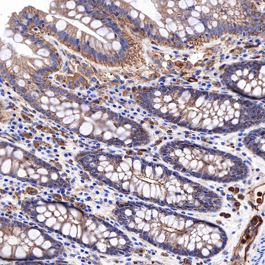
IHC shows positive staining in paraffin-embedded human colon. Anti-α-Actinin-1 antibody was used at 1/2000 dilution, followed by a HRP Polymer for Mouse & Rabbit IgG (ready to use). Counterstained with hematoxylin. Heat mediated antigen retrieval with Tris/EDTA buffer pH9.0 was performed before commencing with IHC staining protocol.
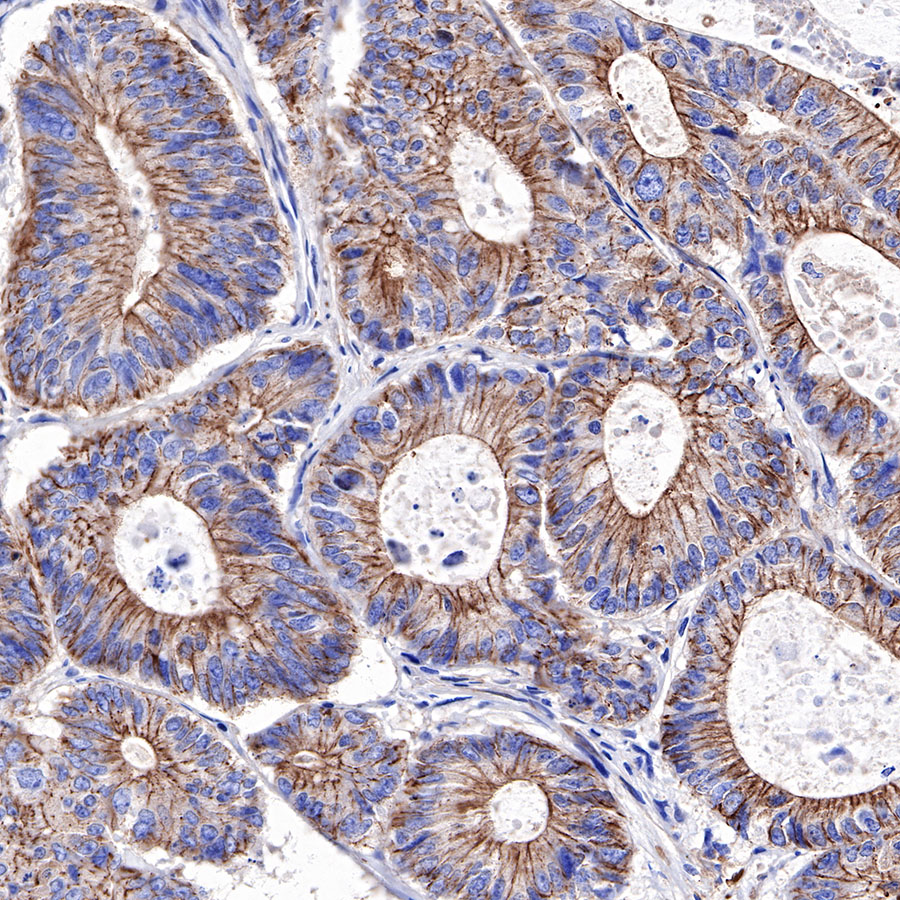
IHC shows positive staining in paraffin-embedded human colon cancer. Anti-α-Actinin-1 antibody was used at 1/2000 dilution, followed by a HRP Polymer for Mouse & Rabbit IgG (ready to use). Counterstained with hematoxylin. Heat mediated antigen retrieval with Tris/EDTA buffer pH9.0 was performed before commencing with IHC staining protocol.
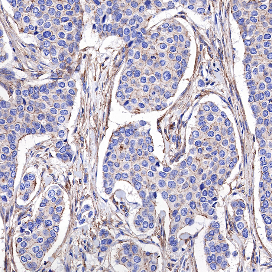
IHC shows positive staining in paraffin-embedded human pancreatic cancer. Anti-α-Actinin-1 antibody was used at 1/2000 dilution, followed by a HRP Polymer for Mouse & Rabbit IgG (ready to use). Counterstained with hematoxylin. Heat mediated antigen retrieval with Tris/EDTA buffer pH9.0 was performed before commencing with IHC staining protocol.
IHC shows positive staining in paraffin-embedded human breast cancer. Anti-α-Actinin-1 antibody was used at 1/2000 dilution, followed by a HRP Polymer for Mouse & Rabbit IgG (ready to use). Counterstained with hematoxylin. Heat mediated antigen retrieval with Tris/EDTA buffer pH9.0 was performed before commencing with IHC staining protocol.
IHC shows positive staining in paraffin-embedded mouse kidney. Anti-α-Actinin-1 antibody was used at 1/2000 dilution, followed by a HRP Polymer for Mouse & Rabbit IgG (ready to use). Counterstained with hematoxylin. Heat mediated antigen retrieval with Tris/EDTA buffer pH9.0 was performed before commencing with IHC staining protocol.
IHC shows positive staining in paraffin-embedded mouse liver. Anti-α-Actinin-1 antibody was used at 1/2000 dilution, followed by a HRP Polymer for Mouse & Rabbit IgG (ready to use). Counterstained with hematoxylin. Heat mediated antigen retrieval with Tris/EDTA buffer pH9.0 was performed before commencing with IHC staining protocol.
IHC shows positive staining in paraffin-embedded rat kidney. Anti-α-Actinin-1 antibody was used at 1/2000 dilution, followed by a HRP Polymer for Mouse & Rabbit IgG (ready to use). Counterstained with hematoxylin. Heat mediated antigen retrieval with Tris/EDTA buffer pH9.0 was performed before commencing with IHC staining protocol.

评论(0)