


 大包装询价
大包装询价 产品介绍 评论(0)
宿主来源
Rabbit抗原名称
pan (alpha) actin分子别名
ACTC1,Alpha-cardiac actin免疫原
Synthetic Peptide细胞定位
Cytoskeleton, CytoplasmAccession
P68032克隆号
SDT-044-17抗体类型
Rabbit mAb应用
IHC-P, ICC, WB, IP, IF反应种属 ?
Hu, Ms, Rt预测反应种属
(反应种属缩写表)Av, Fs, Xe, Sn, SeUr, Dr纯化方式
Protein A浓度
2 mg/ml性状
Liquid缓冲体系
PBS, 40% Glycerol, 0.05%BSA, 0.03% Proclin 300储存条件
12 months from date of receipt / reconstitution, -20 °C as supplied
| 应用 | 稀释度 |
|---|---|
| IP | 1:25 |
| IHC-P | 1:1000 |
| WB | 1:1000 |
| IF | 1:500 |
Actin participates in many important cellular processes, including muscle contraction, cell motility, cell division and cytokinesis, vesicle and organelle movement, cell signaling, and the establishment and maintenance of cell junctions and cell shape. Many of these processes are mediated by extensive and intimate interactions of actin with cellular membranes. In vertebrates, three main groups of actin isoforms, alpha, beta, and gamma have been identified. The alpha actins, found in muscle tissues, are a major constituent of the contractile apparatus. The beta and gamma actins coexist in most cell types as components of the cytoskeleton, and as mediators of internal cell motility.
免疫印迹
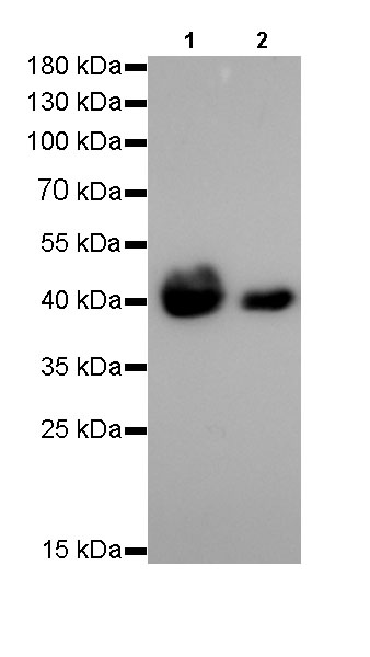
WB result of pan(alpha) actin Rabbit mAb
Primary antibody: pan(alpha) actin Rabbit mAb at 1/1000 dilution
Lane 1: mouse skeletal muscle lysate 20 µg
Lane 2: mouse heart lysate 20 µgSecondary antibody: Goat Anti-Rabbit IgG, (H+L), HRP conjugated at 1/10000 dilution
Predicted MW: 42 kDa
Observed MW: 42 kDa
Exposure time: 6s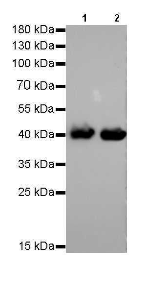
WB result of pan(alpha) actin Rabbit mAb
Primary antibody: pan(alpha) actin Rabbit mAb at 1/1000 dilution
Lane 1: rat skeletal muscle lysate 20 µg
Lane 2: rat heart lysate 20 µgSecondary antibody: Goat Anti-Rabbit IgG, (H+L), HRP conjugated at 1/10000 dilution
Predicted MW: 42 kDa
Observed MW: 42 kDa
Exposure time: 6s
免疫沉淀
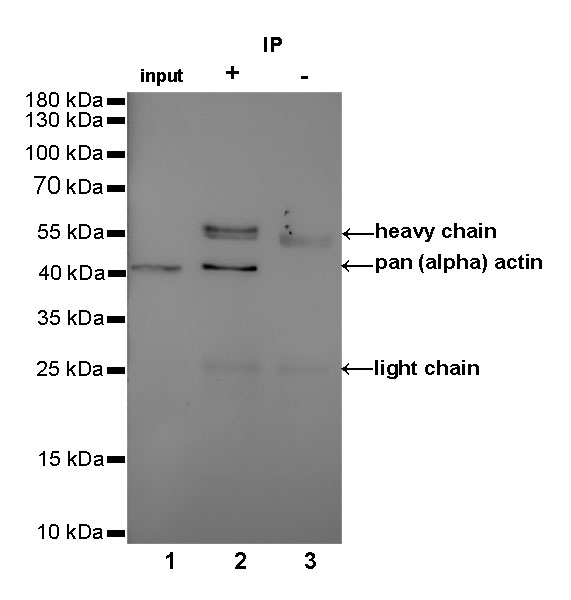
pan (alpha) actin Rabbit mAb at 1/25 dilution (2µg) immunoprecipitating pan (alpha) actin in 0.4mg Mouse skeletal muscle lysate. Western blot was performed on the immunoprecipitate using pan (alpha) actin Rabbit mAb at 1/1000 dilution. Secondary antibody (HRP) for IP was used at 1/400 dilution.
Lane 1: Mouse skeletal muscle lysate 10µg (input)
Lane 2: pan (alpha) actin Rabbit mAb IP in Mouse skeletal muscle lysate
Lane 3: Rabbit monoclonal IgG IP in Mouse skeletal muscle lysate
Predicted MW: 42 kDa
Observed MW: 42 kDa
Exposure time: 180s
免疫组化
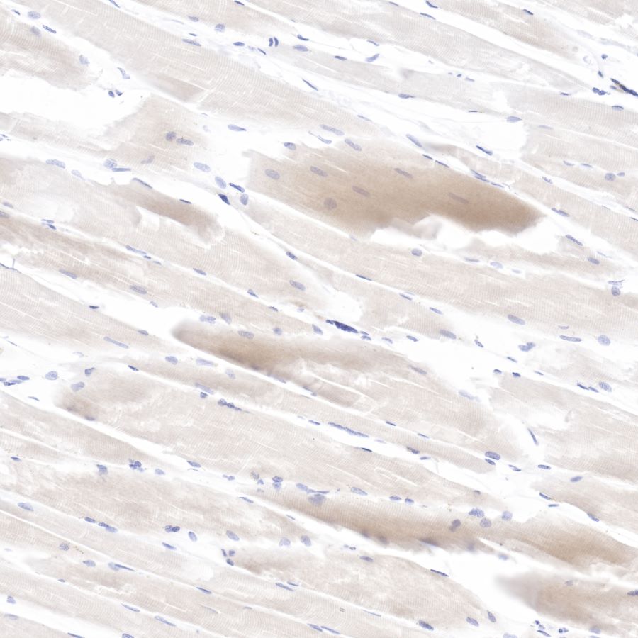
IHC shows positive staining in paraffin-embedded human skeletal muscle.
Anti-pan(alpha) actin antibody was used at 1/1000 dilution, followed by a Goat Anti-Rabbit IgG H&L (HRP) ready to use.
Counterstained with hematoxylin.
Heat mediated antigen retrieval with Tris/EDTA buffer pH9.0 was performed before commencing with IHC staining protocol.
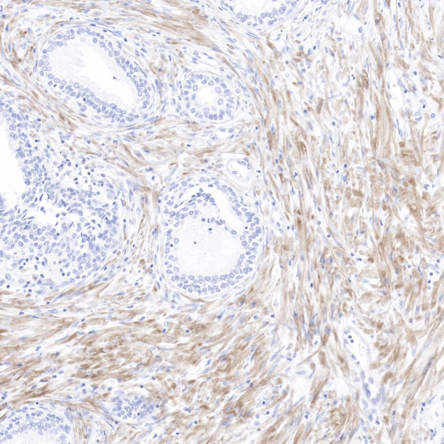
IHC shows positive staining in paraffin-embedded human prostate.
Anti-pan(alpha) actin antibody was used at 1/1000 dilution, followed by a Goat Anti-Rabbit IgG H&L (HRP) ready to use.
Counterstained with hematoxylin.
Heat mediated antigen retrieval with Tris/EDTA buffer pH9.0 was performed before commencing with IHC staining protocol.
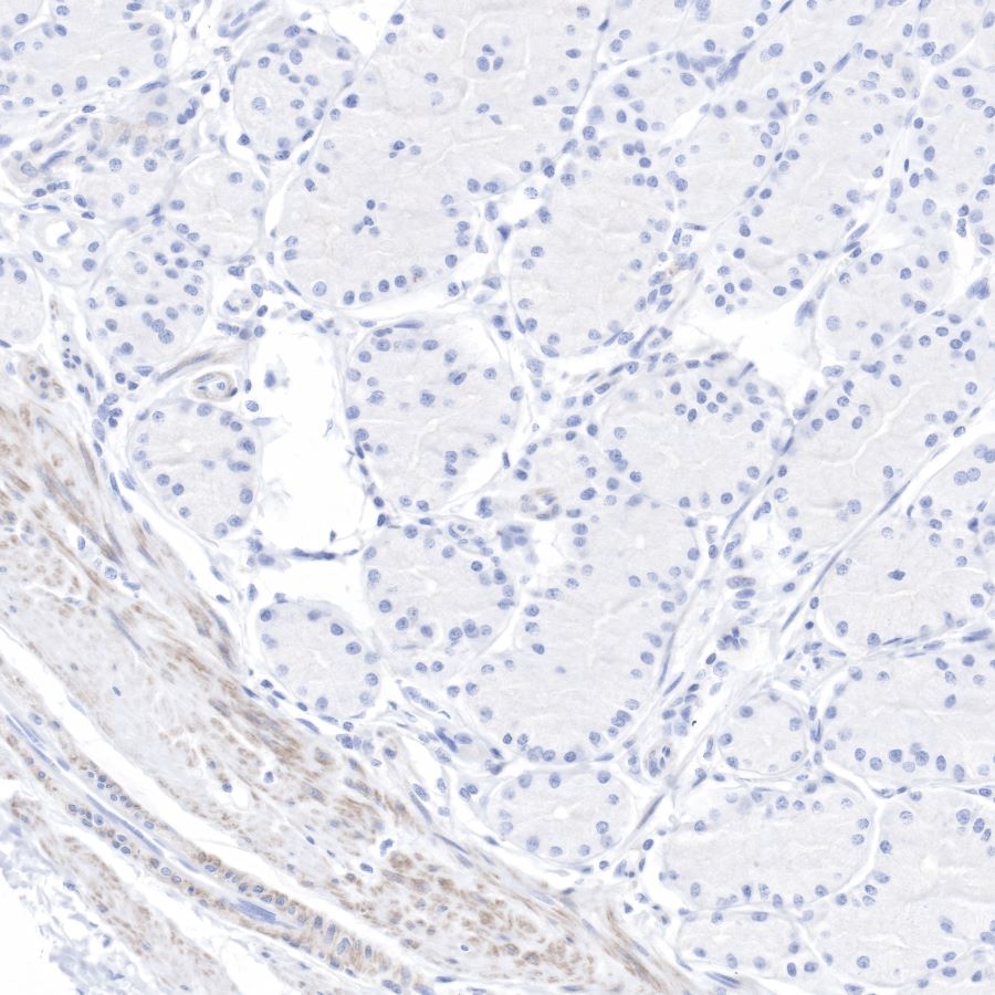
IHC shows positive staining in paraffin-embedded human stomach.
Anti-pan(alpha) actin antibody was used at 1/1000 dilution, followed by a Goat Anti-Rabbit IgG H&L (HRP) ready to use.
Counterstained with hematoxylin.
Heat mediated antigen retrieval with Tris/EDTA buffer pH9.0 was performed before commencing with IHC staining protocol.
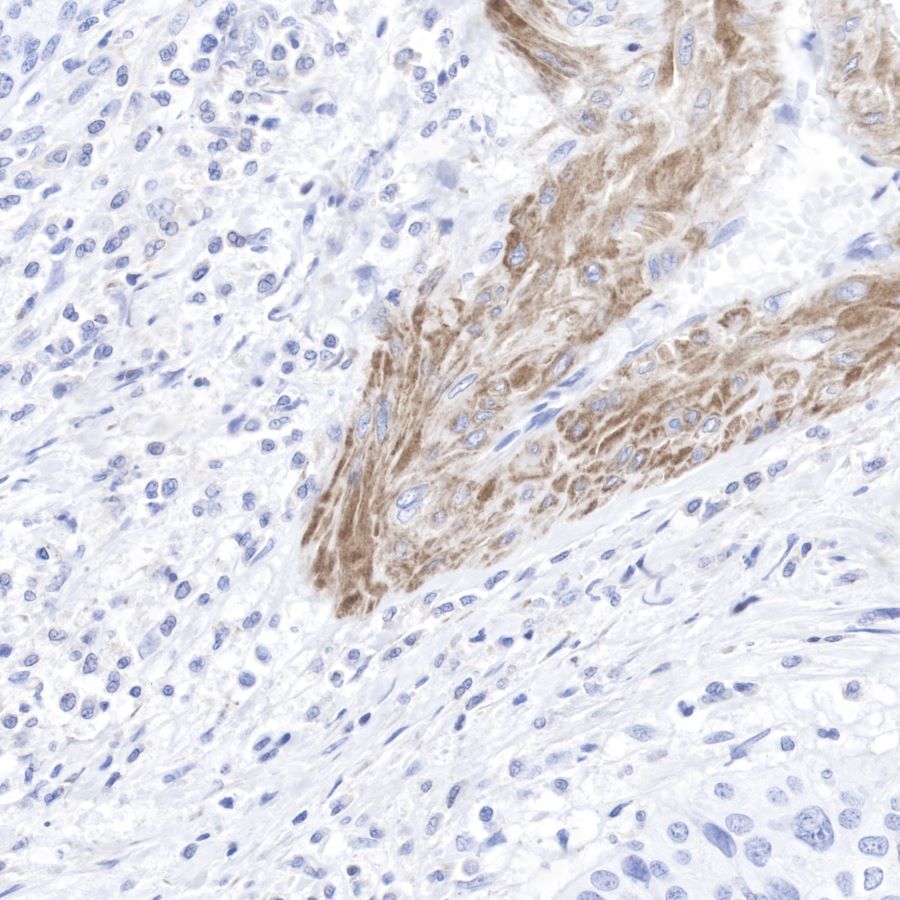
IHC shows positive staining in paraffin-embedded human cervix cancer.
Anti-pan(alpha) actin antibody was used at 1/1000 dilution, followed by a Goat Anti-Rabbit IgG H&L (HRP) ready to use.
Counterstained with hematoxylin.
Heat mediated antigen retrieval with Tris/EDTA buffer pH9.0 was performed before commencing with IHC staining protocol.
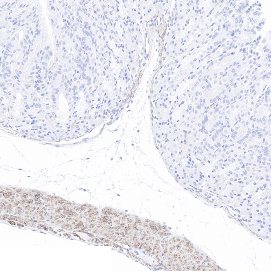
IHC shows positive staining in paraffin-embedded mouse stomach.
Anti-pan(alpha) actin antibody was used at 1/1000 dilution, followed by a Goat Anti-Rabbit IgG H&L (HRP) ready to use.
Counterstained with hematoxylin.
Heat mediated antigen retrieval with Tris/EDTA buffer pH9.0 was performed before commencing with IHC staining protocol.
免疫荧光
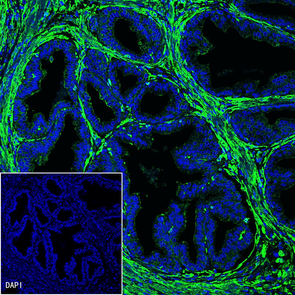
IF shows positive staining in paraffin-embedded human prostate. Anti-pan (alpha) actin antibody was used at 1/500 dilution (Green) and incubated overnight at 4°C. Goat polyclonal Antibody to Rabbit IgG - H&L (Alexa Fluor® 488) was used as secondary antibody at 1/1000 dilution. Counterstained with DAPI (Blue). Heat mediated antigen retrieval with EDTA buffer pH9.0 was performed before commencing with IF staining protocol.
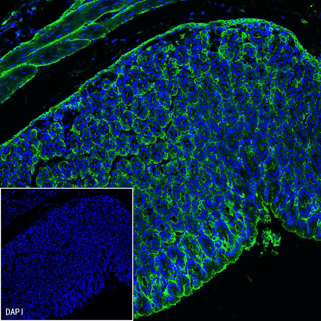
IF shows positive staining in paraffin-embedded mouse stomach. Anti-pan (alpha) actin antibody was used at 1/500 dilution (Green) and incubated overnight at 4°C. Goat polyclonal Antibody to Rabbit IgG - H&L (Alexa Fluor® 488) was used as secondary antibody at 1/1000 dilution. Counterstained with DAPI (Blue). Heat mediated antigen retrieval with EDTA buffer pH9.0 was performed before commencing with IF staining protocol.
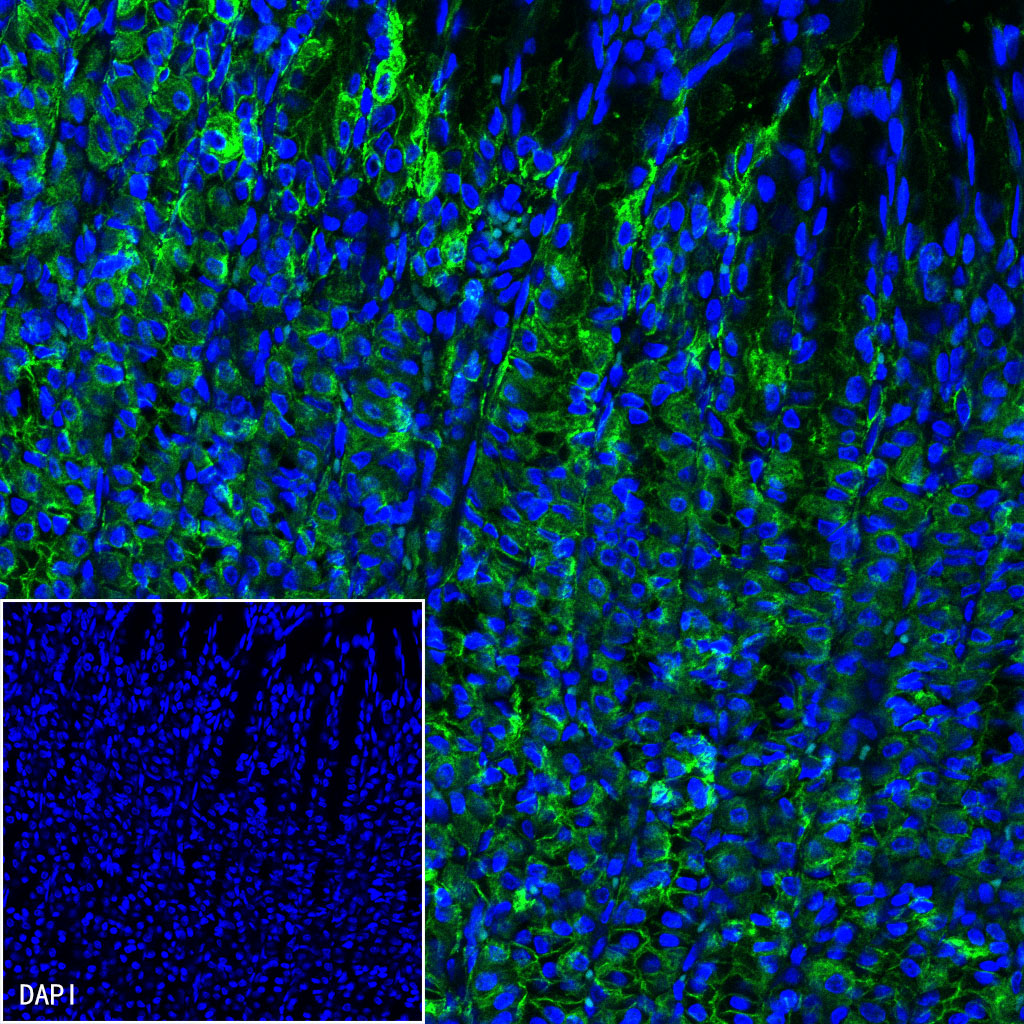
IF shows positive staining in paraffin-embedded rat stomach. Anti-pan (alpha) actin antibody was used at 1/500 dilution (Green) and incubated overnight at 4°C. Goat polyclonal Antibody to Rabbit IgG - H&L (Alexa Fluor® 488) was used as secondary antibody at 1/1000 dilution. Counterstained with DAPI (Blue). Heat mediated antigen retrieval with EDTA buffer pH9.0 was performed before commencing with IF staining protocol.

评论(0)