


 大包装询价
大包装询价 产品介绍 评论(0)
宿主来源
Rabbit抗原名称
Beta 2 microglobulin分子别名
Beta-2-microglobulin form pI 5.3, B2M免疫原
Recombinant Protein细胞定位
Cell Surface, SecretedAccession
P61769克隆号
SDT-096-100抗体类型
Rabbit mAb应用
IHC-P, WB反应种属 ?
Hu, Ms预测反应种属
(反应种属缩写表)Or纯化方式
Protein A浓度
0.25mg/ml性状
Liquid缓冲体系
PBS, 40% Glycerol, 0.05%BSA, 0.03% Proclin 300储存条件
12 months from date of receipt / reconstitution, -20 °C as supplied
| 应用 | 稀释度 |
|---|---|
| IHC-P | 1:1000 |
| WB | 1:500 |
- β2 microglobulin (B2M) is a component of MHC class I molecules. MHC class I molecules have α1, α2, and α3 proteins which are present on all nucleated cells (excluding red blood cells). In humans, the β2 microglobulin protein is encoded by the B2M gene. An additional function is association with the HFE protein, together regulating the expression of hepcidin in the liver which targets the iron transporter ferroportin on the basolateral membrane of enterocytes and cell membrane of macrophages for degradation resulting in decreased iron uptake from food and decreased iron release from recycled red blood cells in the MPS (mononuclear phagocyte system) respectively. Loss of this function causes iron excess and hemochromatosis. In patients on long-term hemodialysis, it can aggregate into amyloid fibers that deposit in joint spaces, a disease, known as dialysis-related amyloidosis. Levels of β2 microglobulin can be elevated in multiple myeloma and lymphoma, though in these cases primary amyloidosis (amyloid light chain) and secondary amyloidosis (amyloid associated protein) are more common.[clarification needed] The normal value of β2 microglobulin is < 2 mg/L.
免疫印迹
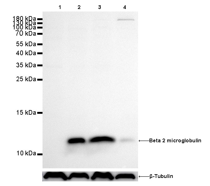
WB result of beta 2 microglobulin Rabbit mAb Primary antibody: beta 2 microglobulin Rabbit mAb at 1/500 dilution
Lane 1: Daudi whole cell lysate 20 µg
Lane 2: Hela whole cell lysate 20 µg
Lane 3: Jurkat whole cell lysate 20 µg
Lane 4: HepG2 whole cell lysate 20 µg
Negative control: Daudi whole cell lysate Secondary antibody: Goat Anti-Rabbit IgG, (H+L), HRP conjugated at 1/10000 dilution
Predicted MW: 12 kDa
Observed MW: 12 kDa
Exposure time: 180s
免疫组化
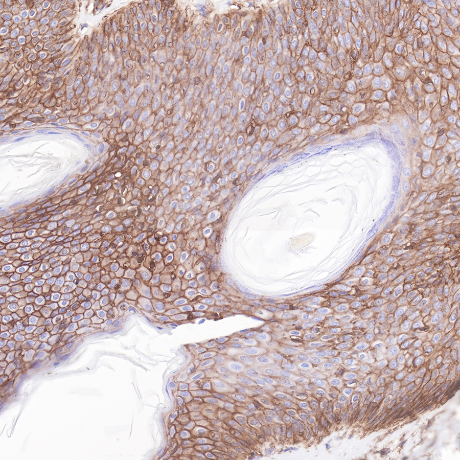
IHC shows positive staining in paraffin-embedded human skin.
Anti-Beta 2 microglobulin antibody was used at 1/1000 dilution, followed by a Goat Anti-Rabbit IgG H&L (HRP) ready to use.
Counterstained with hematoxylin.
Heat mediated antigen retrieval with Tris/EDTA buffer pH9.0 was performed before commencing with IHC staining protocol.
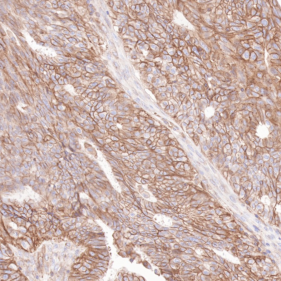
IHC shows positive staining in paraffin-embedded human ovarian cancer.
Anti-Beta 2 microglobulin antibody was used at 1/1000 dilution, followed by a Goat Anti-Rabbit IgG H&L (HRP) ready to use. Counterstained with hematoxylin.
Heat mediated antigen retrieval with Tris/EDTA buffer pH9.0 was performed before commencing with IHC staining protocol.
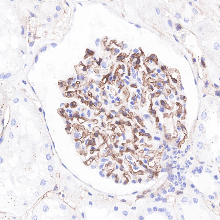
IHC shows positive staining in paraffin-embedded human kidney. Anti-Beta 2 microglobulin antibody was used at 1/1000 dilution, followed by a Goat Anti-Rabbit IgG H&L (HRP) ready to use.
Counterstained with hematoxylin.
Heat mediated antigen retrieval with Tris/EDTA buffer pH9.0 was performed before commencing with IHC staining protocol.
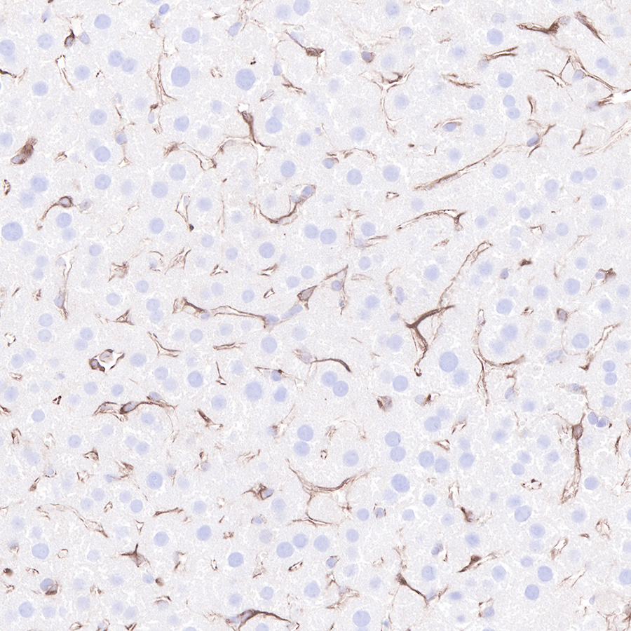
IHC shows positive staining in paraffin-embedded mouse liver.
Anti-Beta 2 microglobulin antibody was used at 1/1000 dilution, followed by a Goat Anti-Rabbit IgG H&L (HRP) ready to use.
Counterstained with hematoxylin.
Heat mediated antigen retrieval with Tris/EDTA buffer pH9.0 was performed before commencing with IHC staining protocol.

评论(0)